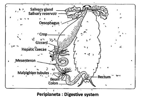
Describe the digestive system of cockroach with the help of a neat labelled diagram.
The digestive system of cockroach consists of an alimentary canal and the associated glands. The preoral cavity, surrounded by the mouth parts, is present in front of the mouth. The hypopharynx divides it into two chambers called cibarium (anterior) and salivarium (posterior).
Alimentary canal: The alimentary canal of cockroach is a long tube and is coiled at some places. It extends between the mouth and the anus, it is divided into three regions, namely, foregut or stomodaeum, midgut or mesenteron, and hindgut or proctodaeum. The forgut and highgut are internally lined by ectoderm. The mesenteron is lined by the endodermal cells.
Foregut of stomodaeum: The foregut includes pharynx, oesophagus crop, and gizzard. It is internally lined by a chitinous cuticle. Mouth opens into the pharynx, which in turn leads into a narrow tubular oesophagus. The oesophagus opens behind into a thin walled, distensible sac called crop. The crop serves as a reservoir for storing food. Its outer surface is covered by a network of tracheae.

Behind the crops there is a thick walled musular proventriculus, or gizzard. The chitinous inner lining of the gizzard has six powerful teeth, which form an efficient grinding apparatus. Behind each tooth is a hairy pad, which bears backwardly directed bristles. Among these plates, food is throughly ground into fine particles. These food particles are filtered by the bristles. The gizzard thus acts both as a grinding mill and also as a sieve. There is a membranous projetion of the gizzard into the mesenteron in the form of a funnel called stomodeal valve. This valve prevents the entry (regurgitation ) of food from the mesenteron back into the gizzard.
Midgut (mesenteron or ventriculus): The midgut is a short and narrow tube behind the gizzard. It is also called mesenteron or ventriculus. Between the ventriculus and the gizzard, arising from ventriculus, there are six to eight finger like diverticula called hepatic caesea. They are helpful in digestion and absorption of the digestion food materials. Ventriculus is functionally divided into an anterior secretory part and a posterior absorptive part.
The secretory part of the ventriculus has many gland cells adn it secretes several enzymes. The bolus of food in teh mesenteron is enveloped by a chitinous and porous membrane called peritrophic membrane, which is secreted by the funnel like stomodeal valve of the gizzard.
Digested food is absorbed into the blood through the peritrophic membrane in the posterior absorptive region of the ventriculus. The peritrophic memberane protects the wall of the ventriculus from hard food particles in the food. The opening of the ventriculus into the hindgut is controlled by a sphincter muscle. It prevents entry of undigested food and uric acid from the hindgut into the midgut.
Hindgut or proctodaeum: The hindgut is a long colled tube, consisting of three regions namely ileum, colon and rectum.It is internaly lined by chitinous cuticle. The ileum that lies behind the mesenteron is a short tube. Six bundles of fine, yellow, blind tubules called maklpighian tubules open into the ileum near the jucntion of mesenteron and ileum. Malpighian tubules are excretory in function. ileum collects uric ancd from the malpighian tubules adn undigested food from the mesenteron. ileum opens behind into a long coiled tube called colon. Colon leads into a short and wide rectum, which opens out through the anus. Retum bears on its inner side six longitudinal chitinous folds called rectal papilae. They are concerned with the reabsorption of water from the undigested food.
Digestive glands: The digestive associated with the alimentary canal of cockroach are salivary glands, hepatic caecae and glandular cells of the mesenteron.
Salivary glands: There is a pair of salivary glands attached to the ventrolateral sides of the crop, one on each side. Each salivary gland has two lobes. Each lobe of salivary gland has many lobules called acini. Each acinus is a group of seccretory cells called zymogencells with a small ductule. The ductules of both the lobes of a salivary gland unite to for a common salivary duct on each side.

The two common salivary ducts are joined toform the median salivary duct. Between the two lobes of a salivary gland of each side is a sac called salivary receptacle that store s saliva. It leads into a receptacular duct, or reservoir duct. The receptacular ducts of both the sides are united to form a common receptacular duct, or common reservoir duct.The median salivary duct opens into the common receptacular duct. Later these two form an efferent salivary duct. The efferent salivary duct opens at the base of the hypopharynx. Acinar cells secrete saliva, which contains starch digesting engymes such as amylase.
Related Playlists
Similar Questions
Describe the digestive system of cockroach with the help of a labelled sketch.
View SolutionDescribe reproductive system of female cockroach with the help of a labelled diagram.
View SolutionExplain oogenesis with the help of a neat labelled diagram
View SolutionDescribe the mechanism of translation with the help of neat and labelled diagram.
View SolutionDescribe functional areas of cerebrum with the help of neat and labelled diagram .
View SolutionDescribe the process of semiconservative replication of DNA with the help of neat and labelled diagram.
View SolutionDescribe the biogas plant with a neat labelled diagram.
View SolutionDescribe the construction and working of a transformer with a neat labelled diagram
View SolutionWith the help of a neat and labelled diagram, describe reflex arc.
View SolutionWith the help of a neat and labelled diagram, describe reflex arc.
View Solution

