Describe human female reproductive system with labelled diagram.Solution in Punjabi
Human Female Repdocutive System. It is concerned with formation of ova, fertilization, foetal development and child-birth. It is also located in the pelvis region. It is formed of a pair of ovaries alongwith their oviducts, uterus, cervix, vagina and the external genitalia. Fallopian tubes, uterus and vagina collectively form the female accessory ducts.
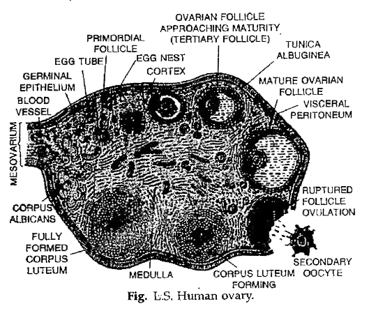
1. Ovaries. Ovaries are the primary sex-organs of female. These are one pair, small sized (2-4 cm x 1.5 cm x 1 cm), almond-shaped structures present in the pelvis, one on either side of uterus. Each ovary is suspended from dorsal abdominal wallby a mesentery called mesovarium. Each ovary is attached to the pelvic wall and uterus by a double fold of peritonerm, ovarian ligament. So ovaries, unlike the testes, are abdominal in position. Each ovary (Fig.) is solid internally and is lined by a cuboidal germinal epithelium. Inner stroma or matrix is differentiated into outer cortex of reticular connective tissue and inner medulla of areolar connective tissue. Outer to ovarian cortex and inner to germinal epithelium, there is a weakly developed connective tissue layer, called tunica albuginea. There are a number of small, oval or rounded developing primary or ovarian follicles in different stages of oogenesis in the medullary region of the ovary. Every month, a primary follicle transforms into mature or Graafian follicle under the stimulation of FSH (Follicular Stimulating Hormone) of anterior pituitary.
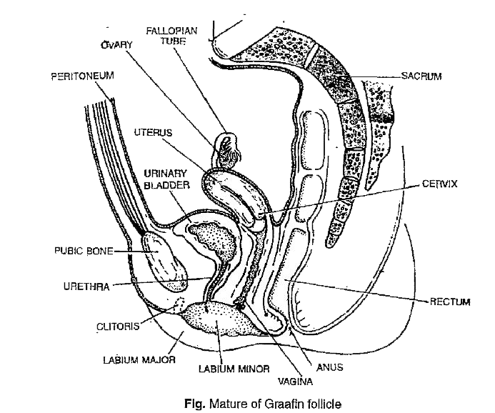
Cortex of ovary may also have yellowish, conical endocrine gland, corpus luteum (yellow body) or a degenerating corpus luteum, corpus albicans (white body). Functions. Ovaries are concerned with formation of ova and secretion of female sex horniones estrogen and progesterone. 2. Fallopian Tubes (Oviducts). These are i long (10-12 cm), ciliated, muscular and tubular« which extend from the periphery of ovaries to uteros Each fallopian tube is differentiated into three parts : (a) Ampulla. It is outer, wide (3 mm in diameter^ funnel-shaped part which opens in abdominal cavity close to ovary by a fimbriated aperture called ostium. Ostium receives the ovum released from the ovary. Tc increase the catching area, it is with a number of finger-like ciliated processes, fimbriae. Its outer expanded and fimbriated part is called infundibulum. (b) Isthmus. It is middle, narrow and ciliated. (c) Uterine part. It is inner and narrow part which opens in the upper part of uterus. Functions. It is involved in the conduction of ovum or zygote towards the uterus by peristalsis and ciliary action. It is also the site of fertilization. 3. Uterus (womb). It is a large (8 cm x 5 cm x 2 cm), hollow, muscular, highly vascular and inverted pear-shaped structure present in the pelvis between the bladder and rectum. It is formed of three parts (Fig.) :
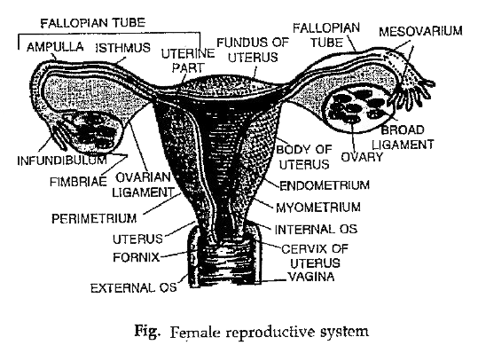
(a) Fundus. It is upper dome-shaped part. (b) Body or Corpus. It is middle and main part of uterus. Its a wall is formed of outer peritoneal layer, perimetrium, middle muscular coat or myometrium of smooth muscle fibres and inner highly vascular and glandular mucosa or endometrium. (c) Cervix. It is lower, narrow part which opens in body of uterus by internal os and in vagina below by external os. The cavity of cervix is called cervical canal and forms a part of birth canal.
Functions. Endometrium of uterus is the site of implantation and foetal growth during pregnancy. It also takes part in placenta formation. Myometrium helps in expelling of the baby during parturition.
4. Vagina. It is a long (7.5 cm), fibro-muscular tube. It extends backward in front of rectum and anal canal from cervix to the vestibule. It is highly vascular tube internally lined by mucous membrane and is raised into transverse folds called vaginal rugae. It finally opens in the vestibule by vaginal orifice.
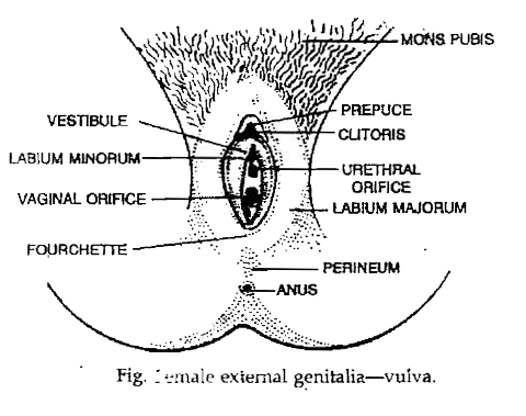
Functions. Vagina acts both as copulation canal as it receives the sperms from penis during copulation and birth canal (Cervical canal + vagina) during parturition. 5. Vulva (Fig.). It is external genitalia of female. It has a depression, the vestibule (or urinogenital sinus), in front of anus. Vestibule has two apertures-upper external urethral orifice of urethra and lower vaginal orifice of vagina. In the virgin female, vaginal orifice is partially covered by a plembranous fold, called hymen, which is often torn during the first coitus (intercourse) or due to vigorous physical activity during horseback riding, bteyeling sudden jerk, etc. but may persist even after coitus. So the presence or absence of hymen is not a reliable indicator of virginity or sexual experience. Vestibule is bounded by two pairs of moist skin folds with sebaceous glands-inner smaller pair called labia minora and outer larger pair called labia majora. Labia minora fuse anteriorly to form a skin fold called prepuce in front of a small erectile organ, the glans clitoridis which is homologous to penis as both are supported by corpora cavernosa. Labia minora also fuse posteriorly to form a membranous fold called fourchette. There is a fleshy elevation above the labia majora and is known as mons pubis which has pubic hair.
Female accessory sex glands
1. Vestibular glands are of two types (a) The lesser vestibular glands (Paraurethral glands or glands of skene) are numerous minute glands that are prsent on either side of the urethral orifice (opening). These glands are homologous to the male prostate and secrete mucus. (b) The greater vestibular glands (Bartholin.s glands) are paired glands, situated one on either side of the vaginal orifice. These glands are homologous to the bulbo-urethral (Cowper.s) glands of male and secrete mucus to lubricate vagina during moting and parturition. 2. Breasts. The breasts are two rounded prominences that lie over the pectoralis major muscles on the front wall of the thorax. They are also present in the male but only in a rudimentary form. In the female, they are undeveloped until puberty. puberty in females they begin to develop under the influence of oestrogen and progesterone hormones. Externally, each breast has a projection, the nipple surrounded by a circular pigmented area of skin (deep pink to light brown) called areola. On the surface of the areola there are numerous sebaceous glands called areolar glands. Internally, the breat consists of the glandular tissue forming mammary glands, the fibrous tissue (connective tissue) and the fatty "or adipose tissue. Mammary glands are modified sweat glands.
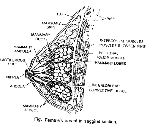
(a) The glandular tissue comprises about 15-20 mammary lobes in each breast. Each lobe is made up of a number of grapelike clusters of milk secreting structures termed alveoli. When milk is produced it passes from the alveoli into the mammary tubules and then into the mammary ducts. Near the nipple, mammary ducts expand to form mammary ampullae ( = lactiferous sinuses) where some milk may be stored before going to lactiferous ducts. Each lactiferous duct typically carries milk from one of the lobes to exterior. (b) The fibrous tissue (connective tissue) supports the alveoli and the ducts, (c) The fatty or adipose tissue is found between the lobes and covers the surface of the gland. The amount of the adipose tissue determines the size of the breasts. Main functions of the mammary glands are secretion and ejection (release) of milk. These functions are called lactation. Lactation is associated with pregnancy and child birth. Milk production is stimulated largely by the hormone prolactin secreted by anterior lobe of the pituitary gland. The ejection of milk is stimulated by the hormone oxytocin, released from the posterior lobe of the pituitary gland. Human milk consists of water and organic and inorganic substances. Its main constituents are fat (fat droplets), casein (milk protein), lactose (milk sugar), mineral salts (sodium, calcium, potassium, phosphorous, etc.) and vitamins. Milk is poor in iron content. Vitamin C is present in very small quantity in milk. The process of milk secretion is regulated by the nervous system. It is. also influenced by the psychic state of the mother. The process of milk production is also influenced by hormones of the pituitary gland (already mentioned), the ovaries and other endocrine glands, A nursing woman secretes 1 to 2 litres of milk per day. Inhibitory Peptide. Milk contains an inhibitory peptide. If the mammary glands are not fully emptied the peptide accumulates and inhibits milk production.
Topper's Solved these Questions
- HUMAN REPRODUCTIONBOOK - BETTER CHOICE PUBLICATIONCHAPTER - HUMAN REPRODUCTIONEXERCISE - SHORT ANSWER TYPE QUESTIONS (MOAT EXPECTED QUESTIONS)
3 Videos
- HUMAN HEALTH AND DISEASEBOOK - BETTER CHOICE PUBLICATIONCHAPTER - HUMAN HEALTH AND DISEASEEXERCISE - LONG ANSWER TYPE QUESTIONS (MOST EXPECTED QUESTIONS)
12 Videos
- MICROBES IN HUMAN WELFAREBOOK - BETTER CHOICE PUBLICATIONCHAPTER - MICROBES IN HUMAN WELFAREEXERCISE - LONG ANSWER TYPE QUESTIONS (MOST EXPECTED QUESTIONS)
2 Videos
Similar Questions
What is role played by luteinizing hormone in males and females?
View SolutionWrite the causative organism, mode of infection, symptoms and preventive measures of the disease pertussis.
View SolutionExplain the role of microbes in the treatment of sewage.
View SolutionDraw a labelled diagram of the reproductive system in a human female.
View SolutionHuman Male and female reproductive system
View SolutionDraw a neat labelled diagram of sectional view of human female reproductive system.
View SolutionDescribe reproductive system of female cockroach with the help of a labelled diagram.
View SolutionDraw a well labelled diagram of sectional view of human female reproductive system.
View SolutionDescribe the human female reproductive system with the help of labelled diagram.
View SolutionDescribe the structure of human female reproductive system with a neat labelled diagram
View SolutionDescribe in brief the female reproductive system of human with diagram.
View SolutionDraw a labelled diagram of a human female reproductive system.
View SolutionDraw a labelled diagram of the reproductive system in a human female.
View SolutionDescribe human female reproductive system with labelled diagram.
View SolutionDescribe in brief the female reproductive system of human with diagram.
View Solution
