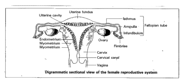
Describe female reproductive system of a woman with the help of a labelled diagram.
The female reproductive system consists of a pair of ovaries along with a pair of oviducts, uterus, vagina and the external genetalia located in the pelvic region. These parts of the system along with a pair of mammary glands are integrated structurally and functionally to support the processes of ovulation, fertilization, pregnancy, birth and child care.

Ovaries Ovaries are the primary female sex organs that produce the female gametes (ova) and several steroid hormones (ovarian hormones). A pair of ovaries is located on each side of lower abdomen. The double layered fold of peritoneum connecting the ovary with the wall of abdominal cavity is known as the meso ovarium.
The Ovaries are covered by a layer of .germinal (ovarian) epithelium. Underneath this layer, there is a dense connective tissue capsule called, .tunica albuginea.. The ovarian stroma is distinctly divided into an outer cortex and an inner medulla. The cortex appears more dense-and-granular-due-to numerous ovarian follicles, The medulla is a loose connective tissue with abundant blood vessels, lymphatic vessels and nerve fibres.
2) Fallopian tubes (oviducts) A Each fallopian tube extends from the periphery of ach ovary to the uterus and-it bears a funnel shaped infundibulum, with finger like projections called .fimbriae., which help in collection of ovum after .ovulation..
The infundibulum leads to a wider part of the oviduct called .ampulla.. The last part of the oviduct, .isthmus) has a narrow lumen and it joins the uterus. Fallopian tube is the site of fertilization. It conducts the ovum or zygote towards the uterus by peristalsis: The fallopian tube is attached to the abdominal wall by a peritoneal fold called .meso salpinx..
3) Uterus The uterus is a single and it is also called womb. It is a large, muscular, highly vascular and inverted pear-shaped structure present in the pelvis between the bladder and rectum. The lower narrow part through which the uterus opens into the vagina is called .cervix.. The cavity of the cervix is called .cervical canal. which along with the vagina forms the .birth canal..
The wall of the uterus has three layers of tissue. The external thin membranous "perimetrium., the middle thick layer of .myometrium. and inner glandulas lining layer called .endometrium. The endometrium undergoes cyclic changes during menstrual cycle while myometrium exhibits strong contractions during parturition.
4) Vagina : The vagina is a large, median, fibromuscular tube that extends from the cervix to the vestibule (the space between labia minora). It is lined by nonkeratinised stratified squamous epithelium. It is highly vascular and opens into the vestibule by the vaginal orifice
5) Vulva Vulva or pudendum refers to the external genitals of the female. The vestibule has two apertures - the upper external urethral orifice of the urethra and the lower vaginal orifice of vagina. Vaginal orifice is covered by a mucous membrane .hymen! vestibule is bound by two pairs of fleshly folds of tissue called inner .labia minora. and outer larger .labia majora.. Clitoris is a sensitive, erectile structure, that lies at the upper junction of the two labia minora above the urethral opening. There is a cushion of fatty tissue covered by skin and pubic hair present above the labia major, called mons pubis.
Accessory reproductive glands of female : These include,
a) Bartholin.s glands : These are two glands located slightly posterior and to the left and right of the opening of the vagina. They secrete mucus to lubricate the vagina and are homologous to the bulbourethral glands of the male reproductive system
b) Skene.s glands : These are located on the anterior wall of vagina, around the lower end of the urethra. They secrete a lubricating fluid when stimulated. The skene.s glands are homologous to the prostate gland of the male reproductive system.
c) Mammary glands: These are paired structures that contain glandular tissue and fat. The alveoli cells present in the mammary lobes of each glandular tissue secrete milk, which is stored in cavities of alveoli. The alveoli open into mammary tubes and then to mammary ducts, from there to mammary ampulla and finally connected to lactiferous duct through which milk is sucked out by the baby. 2.
Related Playlists
- BOOK - VIKRAM PUBLICATION ( ANDHRA PUBLICATION)CHAPTER - HUMAN REPRODUCTIVE SYSTEM EXERCISE - MPORTANT QUESTIONS4videos
HUMAN REPRODUCTIVE SYSTEM
- BOOK - VIKRAM PUBLICATION ( ANDHRA PUBLICATION)CHAPTER - HUMAN REPRODUCTIVE SYSTEM EXERCISE - IMPORTANT QUESTIONS15videos
HUMAN REPRODUCTIVE SYSTEM
- BOOK - VIKRAM PUBLICATION ( ANDHRA PUBLICATION)CHAPTER - HUMAN REPRODUCTIVE SYSTEM EXERCISE - SHORT ANSWER QUESTIONS12videos
HUMAN REPRODUCTIVE SYSTEM
- BOOK - VIKRAM PUBLICATION ( ANDHRA PUBLICATION)CHAPTER - GENETICS EXERCISE - IMPORTANT QUESTIONS13videos
GENETICS
- BOOK - VIKRAM PUBLICATION ( ANDHRA PUBLICATION)CHAPTER - IMMUNE SYSTEM EXERCISE - IMPORTANT QUESTION9videos
IMMUNE SYSTEM
Similar Questions
Describe the female reproductive system.
View SolutionDescribe the female reproductive system.
View SolutionDescribe reproductive system of female cockroach with the help of a labelled diagram.
View SolutionDescribe the male reproductive organs in humans with the help of a labelled diagram.
View SolutionDescribe the human female reproductive system with the help of labelled diagram.
View SolutionWrite a note on human male and female reproductive system with neat labelled diagram
View SolutionDescribe the structure of human female reproductive system with a neat labelled diagram
View SolutionDescribe in brief the female reproductive system of human with diagram.
View SolutionDescribe the human female reproductive system with a labelled diagram .
View SolutionDraw a labelled diagram of a human female reproductive system and explain the function of each organ.
View Solution

