This states that an organisms during its development tends to repeat the evolutionary history of its ancestors Solution in Telugu
. During the development of zygote into a human, a series of events takes place in mother.s womb. They are :
I. Fertillsation
II. Gastrulation
III. Organogenesis
IV. Placenta formation
V. Pregnancy and Parturition.
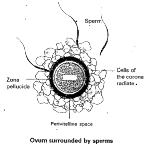
I Fertilisation : It is the formation of zygote by fusing ovum with sperms. Sperm makes it way through corona Sperm radiata and zona pellucida. Acrosin released from acrosome of sperm dissolves zona pellucida of ovum and easiers penetration of sperm into ovum. The entry of sperm, induces the comple - tion of meiosis of ovum. The nuclear union results in the formation of synkaryon (zygotic nucleus).
After fertillsation the events that follow are :

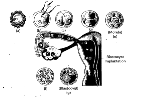
1) Cleavage : After the fertilisation, the first phase of embryonic development in cleavage of zygote as it moves through the isthmus of the oviduct towards the uterus. The daughter cells are called blastomeres.
2) Morulà : Morula is a solid mass of cells and is developed in the fallopian tube and reaches the uterus for further development. The morula passes through a process called compaction, after which the embryo has a superficial flat cell layer and inner cell mass. The outer superficial layer becomes the trophoblast. that serves to attach the embryo to the uterine wall and the inner cell mass constitutes formative cells, which give rise to the .embryo proper., also called the .embryoblast..
3) Blastocyst : Some fluid passes into the morula from uterine cavity thus seperating the inner cell mass from trophoblast. As the quantity of fluid increases, the morula acquires a cyst shape. The cells of trophoblast become flattened and the inner cell mass attaches to the innerside of trophoblast on one side only. The morula now becomes a .blastocyst..
he cavity of the blastocyst is the .blastocoel. or .segmentation cavity. or .primary body cavity.. The side of blastocyst to which the inner cell mass is attached is called .embryonic. or animal pole., while the opposite side is the labembryonic pole.. The cells of the trophoblast above the region of inner cell mass are called .cells of rauber!.
4) Implantation : The zona pellicida around the blastocoel disappears and cells of trophoblast stick to the uterine endometrium. The trophoblast invades endometrium and gets implanted into it, called .interstitial implantation., which starts on the 6th day after fertilisation. After the implantation, the uterine endometrium is differentiated into .decidua..

a) The portion of the decidua where the placenta is to be formed is called the .decidua basalis.
b) The part of the decidua that seperates the embryo from the uterine lumen is called the .decidua capsularis..
c) The part lining the rest of the uterine cavity is called the decidua perietalis.
At the end of pregnancy the decidua is shed off, along with the placenta and membranes.
5) Formation of Bilaminar Embryonic disc : The inner cell mass of blastocyst forms into a disc called .embryonic disc.. Then the .cells of Rauber. disapearts, some,cells get seperated by delamination and eventually forms a layer of cells, that further develops into the hypoblast, which is the future extra embryonic endoderm. The remaining part of the disc. is called .epiblast.. Now the embryonic disc is called .bilaminar embryonic disc..
The hypoblast layer below the trophoblast encloses a cavity called .yolk sac. or .umblical vesicle.. Gradually the embryonic disc becomes oval.
II. Gastrulation : Gastrulation involves proliferation, differentiation of movement of cells with in the embryo. Along the longitudinal axis of the embryonic disc, a primitive streak is formed. A longitudinal furrow, .primitive groove. forms along the middle of primitive streak. On either side of it are the primitive folds.. Anteriorly primitive streak has a shallow primitive pit. The thickened part of streak is called .primitive knot. or .primitive node. or .Hansen.s node
1) Trilaminar Embryo : The furture epidermal cells from epiblast, forms the endoderm, of embryo. The remaining epiblast is known as .ectoderm.. The invasion of epiblast cells into space between the epiblast and hypoblast is called gastrulation. The process of gastrulation converts the bilaminar embryonic disc to trilaminar embryonic disc.
2) Extrà embryonic membranes : Now four extra embryonic or foetal membranes are formed. They are chorion, amnion, allantios and yolk sac.
(a) Between the amnion and the embryo, there is an amniotic cavity filled with aminiotic fluid, that acts as shock absorber and also prevents embryo from shock:
b) Allantios and chorion, are fused to form .chorio allantoic membrane. which constitute placenta.
(C) Yolk sac encloses a fluid cavity, it has no nutritive value
III. Organogenesis : In involves series of stages.
1) Formation of Notochord and Neural tube : The chora mesodermal cells converge and involute through Hensen.s node and extends forward as .notochordal process.. This later transforms into a solid rod - .notochord.. The notochord mięsoderm induces the overlying endodermal cells to form neural cells which further changes into a neural tube by a process called neurlation.
2) Differentiation of Mesoderm and Formation of Coelom : The longitudinal column of mesoderm adjacent to neural tube on either side is called .epimere. and the mesoderm around the gut is .hypomete.. The mesoderm in between these two is .mesomers.. The somites. of epimere differentiate into myotome, sclerotome and dermatome.
(a) Sclerotome - forms the vertebral column
(b) Dermatome.- forms the dermis of the skin
(c) Myotome - forms the voluntary muscles of the body
The hypomere splits into outer somatic and inner splanchnic mesodermal layers: Intra embryonic coelom is formed in between these two layers, which given rise to pericardial, pleural and peritoneal cavities:
IV. Placenta famation : The chorionic villi and’uterine tissue interdigitate with each other to form a structural and functional unit called .placenta. between the foetus and the mother. The maternal and foetal blood are seperated by placental membrane.
Functions of Placenta :
(a) Supplies O2 and nutrients to the embryo.
(b) Removes CO2 and waste materials from embryo.
(c ) Progesterone secreted by it essential for maintenance of pregnancy
(d) Oestrogen secreted by it promotes uterine growth.
(e) hCG produced is used as a test to detect pregnancy.
(f) Somato mammotropin increases glucose levels of plasma.
V. Pregnancy and Parturition :
Pregnancy : It is the intra uterine development of embryo and foetus. In humans it is 266 days (38 weeks) from the fertilization of egg.
Events during pregnancy : Human gestation can be divided into 3 trimesters of three months each. The events are:
One month - Embryo.s heart is formed
Second month - Foetus develops limbs and digits
Third month - Major organs and formed
Fifth month - First movements and appearance of hair and head.
Six months - Body is covered with fine hair, eye lids separate and eye lashes are formed.
Nine months - Foetus is fully developed.
Parturition: The process of delivery of foetus after labour is called parturition and is favoured by hormone oxytocin that causes stronger uterine contractions. This leads to the expulsion of baby out of the uterus through the birth canal.
Topper's Solved these Questions
- HUMAN REPRODUCTIVE SYSTEM BOOK - VIKRAM PUBLICATION ( ANDHRA PUBLICATION)CHAPTER - HUMAN REPRODUCTIVE SYSTEM EXERCISE - MPORTANT QUESTIONS
4 Videos
- HUMAN REPRODUCTIVE SYSTEM BOOK - VIKRAM PUBLICATION ( ANDHRA PUBLICATION)CHAPTER - HUMAN REPRODUCTIVE SYSTEM EXERCISE - IMPORTANT QUESTIONS
15 Videos
- HUMAN REPRODUCTIVE SYSTEM BOOK - VIKRAM PUBLICATION ( ANDHRA PUBLICATION)CHAPTER - HUMAN REPRODUCTIVE SYSTEM EXERCISE - SHORT ANSWER QUESTIONS
12 Videos
- GENETICS BOOK - VIKRAM PUBLICATION ( ANDHRA PUBLICATION)CHAPTER - GENETICS EXERCISE - IMPORTANT QUESTIONS
13 Videos
- IMMUNE SYSTEM BOOK - VIKRAM PUBLICATION ( ANDHRA PUBLICATION)CHAPTER - IMMUNE SYSTEM EXERCISE - IMPORTANT QUESTION
9 Videos
Similar Questions
Testes of man occur
View SolutionDefine spermiogenesis and spermiation.
View SolutionTestes of man occur
View SolutionWhat is Pleiotropy?
View SolutionDefine the terms immunity and immune system.
View SolutionName the law that states 'Embryonic Development of an animal repeats the embryonic stages of ancestors ?
View SolutionThe evolutionary history of an organism is known as
View SolutionEvolutionary history of an organism is called
View SolutionEvolutionary history of an organism is called
View SolutionThis states that an organisms during its development tends to repeat the evolutionary history of its ancestors
View SolutionEvolutionary history of an organism is known as
View SolutionEvolutionary history of an organism is known as
View SolutionEvolutionary history of an organism is known as:
View Solutionकिसी जीव के विकासीय इतिहास को क्या कहते हैं?
View SolutionEvolutionary history of an organism is called
View SolutionThe evolutionary history of an organism is known as
View Solution