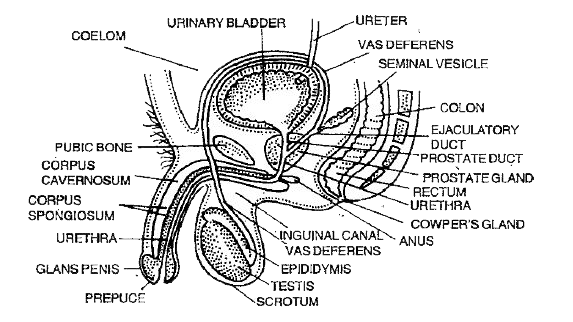
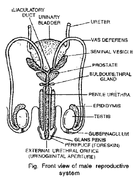
Draw a label, explain the male reproductive system of man. Solution in Punjabi
The male reproductive system is composed of testes together with accessory ducts and structures including epididymis, vas deferens, ejaculatory duct, the distal portion of urethra, the penis and several glands.
1. Testes. There is a pair of testes which lie outside the abdominal cavity in a thin pouch of skin called scrotum. Each testis is oval shaped, pinkish coloured, measures about 4 cm, 3 cm and 2.5 cm in length, thickness and width respectively. The testes are attached to the scrotum at its flat, posterior surface by a small, thick, white fibrous gubernaculum and spermatic cord. The latter is formed of cremaster muscles and connective tissues. The cavity of scrotum remains connected to the abdominal cavity through inguinal canal. The scrotum is thermoregulatory in its function and maintains the temperature of the testes lower by 2 to 3∘C than the body temperature which is favourable for spermatogenesis. The FSH is secreted by anterior pituitary which promotes the descent of testes into the scrotum.
2. Prostate gland. It is the largest auxiliary gland with chestnut like shape situated around the Ist part of the urethra below the urinary bladder. It is surrounded by a thin and dense capsule of fibrous connective tissue and muscle fibres which provide firm palpation to it. It secretes milky, thin and alkaline fluid containing citric acid, bicarbonate ions, lipids and acid phosphatase that gives characteristic, seminal order and alkalinity to the ejaculate. It increases the motility of the sperms and neutralizes the acidity of urine. It constitutes about 5-30% of the ejaculate.
3. Cowper.s glands (Bulbourethral glands). These are paired glands situated beneath the bladder and on each side of the urethra into which their ducts open. They are about the size of pea seed and form the floor of the pelvis. They secrete, clear, white, viscous, alkaline, mucoid lubricant that neutralize the activity of acidic female vaginal secretions and increase the mobility and survival potentiality of sperms in the genital tract of female.

4. Epididymes. The epididymis is a mass of long narrow closely coiled tubule which lies along the inner side of each testis. At the anterior end of the testis it is called caput epididymis, in which the vasa efferentia open. The middle part of the eipdidymis is known as corpus epididymis. The posterior end of the epididymis is called as cauda epididymis. The epididymis stores the sperms and also secretes a fluid which is considered to nourish the sperms.
5. Vasa deferentia. A vas deferens emerges from thie cauda epididymis on each side and leaves the scrotal sac and enters the abdominal cavity through the inguinal canal. The vas deferens loops over the urinary bladder where it is joined by duct from the seminal vesicle to form the ejaculatory duct. Vasa deferentia carry sperms. Rete testis, vasa efferentia, epididymes and vasa deferentia are called the male sex accessory ducts. These ducts store and transport the sperms from the testis to the outside through urethra.
6. Ejaculatory ducts. The ejaculatory ducts are two short tubes each formed by the union of the duct from a seminal vesicle and a vas deferens. They pass through the prostate gland and join the prostatic part of the urethra. The ejaculatory ducts are composed of the fibrous, muscular and columnar epithelial tissue. Ejaculatory ducts carry sperms and secretion of seminal vesicles.
7. Urethra. The male urethra provides a common pathway for the flow of urine called semen. It is much longer in male than in the female, measuring about 20 cm. The urethra includes three parts (i) The first part is surrounded by the prostate gland and is called the prostatic urethra which arises from the urinary bladder and carries urine only, (ii) The second part is the membranous urethra which is situated behind the lower part of the pubic symphysis. The membranous urethra is the smallest urethra, (iii) The third part is the penile urethra which is situated in the penis. There are two urethral sphincters. The internal sphincter consists of smooth muscle fibres situated at the neck of the bladder above the prostate gland. The external sphincter consists of striated muscle fibres surrounding the membranous part of the urethra. Membranous urethra and penile urethra carry both urine and semen. External opening of the urethra is called urethral meatus.
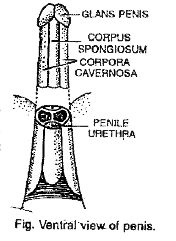
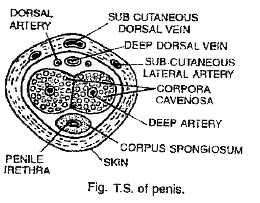
8. Penis. The penis is male genitalia (male copulatory organ). At the tip of the glans penis is the slit like opening called the external urethral orifice or urinogenital aperture. The penis in addition to conducting urine from the body, transfers semen into reproductive tract of the female during sexual intercourse. The penis contains three cylindrical masses of erectile tissue-two dorsal corpora cavernosa and one ventral corpus spongiosum. These bodies are surrounded by fibrous tissue. The corpus spongiosum which contains the penile urethra, is enlarged end of the penis to form the glans penis. The glans penis is covered by loose fold of skin, the prepuce or foreskin. During sexual arousal the three bundles of tissue in the penis become engorged with blood. The penis carries both urine: and semen.

9. Male Accessory glands. Ventral view of penis. (i) The seminal vesicles are one pair of sac like structures near the base of the bladder. Their ducts join the vasa deferentia to form the ejacula-tory ducts. They produce an alkaline secretion which forms 60% of the volume of semen. Seminal fluid pH . is 7.4. The secretion of the seminal vesicles contains fructose, hormone like prostaglandins, and clotting proteins that are different from those in blood. The fructose is a source of energy for the sperm. The prostaglandins stimulate uterine contractions and thus may help the sperm to be moved towards the female.s oviducts, where fertilization takes place. The clotting proteins help semen coagulate after ejaculation. Alkaline nature of the seminal fluid helps to neutralize the acidic environment of the male urethra as well as that of female reproductive tract which otherwise would inactivate and kill sperms. Fructose, which is produced by the seminal vesicles, is not present anywhere else in the body, provides a a forensic test for rape. The presence of fructose, in the female.s genital tract confirms sexual intercourse.

(iii) Prostate gland. It is the largest auxiliary gland with chestnut like shape situated around the 1st part of the urethra below the urinary bladder. It is surrounded by a thin and dense capsule of fibrous connective tissue and muscle fibres which provide firm palpation to it. It secretes milky, thin and alkaline fluid containing citric acid, bicarbonate ions, lipids and acid phosphatase that gives characteristic seminal order and alkalinity to the ejaculate. It increases the motility of the sperms and neutralizes the acidity of urine. It constitutes about 5-30% of the ejaculate.
 (iii) Cowper.s glands (Bulbourethral glands). These are paired glands situated beneath the bladder and on each side of the urethra into which their ducts open. They are about the size of pea seed and form the floor of the pelvis. They secrete, clear, white, viscous, alkaline, mucoid lubricant that neutralize the activity of acidic female vaginal secretions and increase the mobility and survival potentiality of sperms in the genital tract of female.
Topper's Solved these Questions
- HUMAN REPRODUCTIONBook:BETTER CHOICE PUBLICATIONChapter:HUMAN REPRODUCTIONExercise:SHORT ANSWER TYPE QUESTIONS (MOAT EXPECTED QUESTIONS)
Explore 3 Videos
- HUMAN HEALTH AND DISEASEBook:BETTER CHOICE PUBLICATIONChapter:HUMAN HEALTH AND DISEASEExercise:LONG ANSWER TYPE QUESTIONS (MOST EXPECTED QUESTIONS)
Explore 12 Videos
- MICROBES IN HUMAN WELFAREBook:BETTER CHOICE PUBLICATIONChapter:MICROBES IN HUMAN WELFAREExercise:LONG ANSWER TYPE QUESTIONS (MOST EXPECTED QUESTIONS)
Explore 2 Videos
Similar Questions
What is role played by luteinizing hormone in males and females?
View SolutionWrite the causative organism, mode of infection, symptoms and preventive measures of the disease pertussis.
View SolutionExplain the role of microbes in the treatment of sewage.
View SolutionDraw a labeled diagram of male reproductive system.
View SolutionDraw a labeled diagram of male reproductive system.
View SolutionDraw the male reproductive system of frog & label the parts.
View SolutionExplain the male reproductive system of rabbit with a labelled diagram.
View SolutionDraw a neat labelled diagram of human male reproductive system.
View SolutionDraw a labelled diagram of male reproductive system.
View SolutionExplain the male reproductive system of rabbit with a labelled diagram.
View SolutionDraw a labelled diagram of male reproductive system.
View SolutionDraw a labelled diagram of a human male reproductive system.
View SolutionDraw a labeled diagram of male reproductive system.
View Solutionमेंढक के नर जनन तंत्र का स्वच्छ एवं साफ-साफ एवं अच्छी तरह लेबल किया गया आरेख बनाइए।
View SolutionDraw a well labelled diagram of male reproductive system of man.
View Solution