Describe the structure and life cycle of Ascaris lumbricoides with the help of a neat and labelled diagram.Solution in Telugu
Ascaris lumbricoides is commonly called the common round worin. It lives in the small intestine of man, more frequently in children. It is cosmpolitan in distribution, Mode of infection is through contaminated food and water. Infective stage is the embryonated egg with the 2nd stage rhabditiform larva.
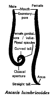
Structure , Sexes are separate and the sexual dimorphism is distinct. In both males and females the body is elongated and cylindrical. Mouth is present at the extreme anterior end and is surrounded by three chitinous lips close to the mouth. Mid ventrally there is a small aperture called excretory pore.
Male : It has a curved posterior and which is considered the tail. The posterior end possesses a cloacal aperture and a pair of equalsized chitinous pineal spicules or pineal setae which serve to transfer the sperms during copulation.
Female : It has a straight posterior end, the tail. The female genital pore or vulva is present mid ventrally at about one third the length from mouth. The anus is present a little in front of the tail end.
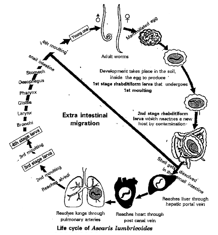
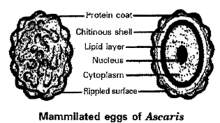
Life history : Copulation takes place in the small intestine of man. After copulation, the female releases approximately two lakh eggs per day. Each egg is surrounded by a protein coat with rippled surface. Hence the eggs of Ascaris are described as mammillated eggs. The protein coat is followed by

a chitinous shell and a lipid layer internally. These eggs come out along with faecal matter. In the moist soil, development takes place inside the egg so that the 16 stage rhabditiform larva is produced. It undergoes the 1" moulting and becomes the 2nd stage rhabditiform larva which is considered the stage infective to man. They reach the alimentary canal of man through contaminated food and water.
In the small intestine, the shell gets dissolved so that the 2nd Male Female stage larva is released. Now it undergoes extra intestinal migration. Mouth First it reaches the liver through the hepatic portal vein. From there it Excretory pore reaches the heart through the post caval vein. It goes to the lungs through the pulmonary arteries. In the alveoli of lungs it undergoes the 2nd moulting to produce the 3rd stage larva. It undergoes the 3rd moulting female genitaloore / vulva so that the 4th stage larva is produced in the alveoli only. It leaves the Pineal spicules alveoli and reaches the small intestine again through bronchi, trachea, Curved tall larynx, glottis, pharynx, oesophagus and stomach. In the small intestine.. It under goes the 4th and final moulting to become a young one which attains sexual maturity with in 8 to 10 weeks.


Topper's Solved these Questions
- BIOLOGY IN HUMAN WELFARE Book:VIKRAM PUBLICATION ( ANDHRA PUBLICATION)Chapter:BIOLOGY IN HUMAN WELFARE Exercise:IMPORTANT QUESTIONS
Explore 10 Videos
- BIOLOGY IN HUMAN WELFARE Book:VIKRAM PUBLICATION ( ANDHRA PUBLICATION)Chapter:BIOLOGY IN HUMAN WELFARE Exercise:SHORT ANSWER TYPE QUESTIONS
Explore 19 Videos
- BIOLOGICAL CLASSIFICATION Book:VIKRAM PUBLICATION ( ANDHRA PUBLICATION)Chapter:BIOLOGICAL CLASSIFICATION Exercise:IMPORTANT QUESTIONS
Explore 7 Videos
- BOARD MODEL PAPER Book:VIKRAM PUBLICATION ( ANDHRA PUBLICATION)Chapter:BOARD MODEL PAPER Exercise:SECTION - C
Explore 3 Videos
Similar Questions
What is hyper parasite ? Mention the name of one hyper parasite.
View SolutionWhat is the need for parasites to develop special adarptations ? Mention some special adaptations developed by the parasites.
View SolutionHow are viroids different from viruses?
View SolutionDescribe the life cycle of plasmodium vivax in mosquito ?
View Solutionఆస్కారిస్ లుంబ్రికాయిడిస్ నిర్మాణాన్ని జీవితచక్ర పటాలతో వివరించండి.
View SolutionDescribe the structure of nervous tissue with the help of a neat labelled diagram.
View SolutionDescribe the structure of anatropous ovule with the help of neat and labelled diagram.
View SolutionWith the help of a neat labelled diagram describe the structure of a chromosome.
View SolutionDraw and label the life cycle of Ascaris lumbricoides.
View SolutionWith the help of a neat labelled diagram, describe the internal structure of dorsiventral leaf. Draw neat labelled diagram: T.S. of Dicot leaf.
View SolutionWith the help of a neat labelled diagram, describe the location and structure of striated muscle fibre.
View SolutionWith the help of a neat labelled diagram, describe the location and structure of nonstriated muscles.
View SolutionDescribe the structure of gizzard of cockroach with the help of neat labelled diagram.
View SolutionWith the help of neat labeled diagram describe the internal structure of human heart.
View SolutionExplain the structure and life cycle of Entamoeba histolytica with the help of neat and labelled diagrams?
View Solution

