Explain the structure and life cycle of Entamoeba histolytica with the help of neat and labelled diagrams?Solution in Telugu
Entamoeba histolytica (Gr. entos - within : amoiba - change histos- tissues, lysis - dissolve) is a microscopic and monogenetic parasite that inhabits the large intestine and causes amoebic dysentery or amoebiasis in man.
It is cosmopolitan in distribution but more comion in the tropical and subtropical regions of the world. It is common in the people of rural and densely urban areas wherever the hygienic conditions are poor.
Structure : Entamoaba histrolytica passes through three stages in its life cycle namely
i) Trophozoite stage
ii) Precystic stage
iii) Cystic stage

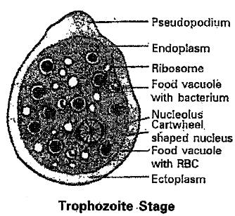
i) Trophozoit stage.: It is the most active, motile, Endoplasm Ribosome feeding and pathogenic stage that lives in the mucosa and Food vacuole sub mucosa membrane of the large intestine. It moves with with bacterium Nucleolus the help of a lobopodium which is produced anteriorly. The Cartwheel shaped nucleus body of the trophozoite is surrounded by plasma- lemma. Food vacuole Its cytoplasm is differentiated into outer clear, viscous non with RBC Ectoplasm granular ectoplasm and inner.fluid ke, granular endoplasm. Ribosomes, food vacuoles and vesicular, cartwhool shape Trophozoite Stage nucleus are present in the endoplasm. The absence of mitochondria indicates the.obligate anaerobic nature of Entamoeba histolytica. It produces the proteolytic enzyme called histolysin due to which the species name histolytica was assigned to it. Due to the effect of thism enzyme the mucosa and submucosa of the gut wall are dissolved releasing some amount of blood, tissue debris which are ingested by the trophozoites. Hence the food vacuoles are with erythrocytes fragments of epithelial cells and bacteria. Presence of RBC in food vacuoles and cartwheel shaped nucleus are the characteristic features of the trophozoites of Entamoeba histlytica.
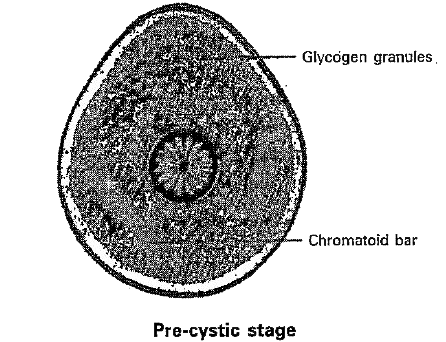
ii) Precystic stage : It is the rion - feeding and non - pathogenic stage of Entamoeba histolytica that is found in the lumen of large intenstine.
It is a small, spherical or oval non-motile form. The cytoplasm of the precystic stage stores glycogen granules and chromotoid bars (made of ribonucleo protein) which act as reserve food.

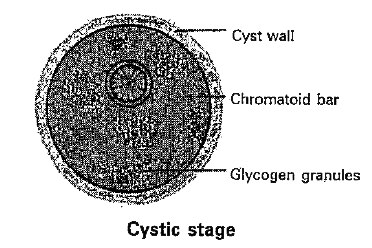
iii) It is round in shape and is surrounded by a thin, delicate and highlt resistant cyst wall. it is found in the lumen of the large intestine. The process of development of cyst wall is called. Encystation. Which is a means of tide over the unfavurable conditions that the parasite is going to encounter while passing to a new host. Soon after the .encystation, the nucleus undergoes two successive Glycogen granules mitotic divisions to form four daughter nuclei. This type of cystic stage is called tetranucleate cyst or mature cyst Cystic stage which is the stage infective to man.

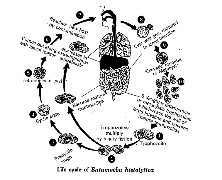
Life cycle : The trophozoites undergo binary fissions in the wall of the large intestine and produce a number of daughter entamoeba. They feed upon the bacteria and the host.s tissue elements, grow in size and again multiply. After repeated binary fission some of the young ones enter of the lumen of the large intestine and transform into precystio stages. Here, the precystic stages transform into cystic stages which in turn develop into tetranucleate cysts. The entire process is completed only in a few hours. These tetranucleate cysts come out along with the faecal matter and can remain alive for about 10 days. The cyst reach new host through contaiminated food and water. In the small intestine of a new human host the cyst wall gets reptured releasing the tetranucleate amoebae. Sach tetranucleate excystic amoebae are called metacysts.
The four nuclei of the metacyst undergo mitotic divisions and produce eight nuclei. Each nucleus gets a bit of the cytoplasm and thus eight daughter entamoebae or metacystic trophozoites are produced. These young ones develop into feeding stages called trophozoites. They invade the mucous membrane of the large intestine and grow into mature trophozoites.

Topper's Solved these Questions
- BIOLOGY IN HUMAN WELFARE BOOK - VIKRAM PUBLICATION ( ANDHRA PUBLICATION)CHAPTER - BIOLOGY IN HUMAN WELFARE EXERCISE - IMPORTANT QUESTIONS
10 Videos
- BIOLOGY IN HUMAN WELFARE BOOK - VIKRAM PUBLICATION ( ANDHRA PUBLICATION)CHAPTER - BIOLOGY IN HUMAN WELFARE EXERCISE - SHORT ANSWER TYPE QUESTIONS
19 Videos
- BIOLOGICAL CLASSIFICATION BOOK - VIKRAM PUBLICATION ( ANDHRA PUBLICATION)CHAPTER - BIOLOGICAL CLASSIFICATION EXERCISE - IMPORTANT QUESTIONS
7 Videos
- BOARD MODEL PAPER BOOK - VIKRAM PUBLICATION ( ANDHRA PUBLICATION)CHAPTER - BOARD MODEL PAPER EXERCISE - SECTION - C
3 Videos
Similar Questions
What is hyper parasite ? Mention the name of one hyper parasite.
View SolutionWhat is the need for parasites to develop special adarptations ? Mention some special adaptations developed by the parasites.
View SolutionHow are viroids different from viruses?
View SolutionDescribe the life cycle of plasmodium vivax in mosquito ?
View SolutionWith the help of a neat labeled diagram, explain structure and any one function of collenchyma.
View Solutionఎంటమీబా హిస్టోలైటికా నిర్మాణాన్ని, జీవిత చక్రాన్ని వివరించండి. పటం గీసి, భాగాలను గుర్తించండి.
View SolutionExplain the structure of mature female gametophyte with the help of a neat labelled diagram.
View SolutionWith the help of a neat labelled diagram describe the structure of a chromosome.
View SolutionSketch and Label-Life cycle of Entamoeba histolytica.
View SolutionExplain the structure of prokaryotic cell with the help of neat labelled diagram.
View SolutionWith the help of neat labelled diagram explain the structure of nucleus.
View SolutionWith the help of neat labeled diagram explain single circulation.
View SolutionShow the life cycle of Entamoeba histolytica with only diagrammatically labeled diagrams (description unnecessary). Discuss the pathogenicity of Entamoeba and the main measures to prevent its infection.
View SolutionDraw neat labelled diagrams.
Haplontic life cycle
View SolutionExplain the life cycle of Silkworm with neat labelled diagram.
View Solution
