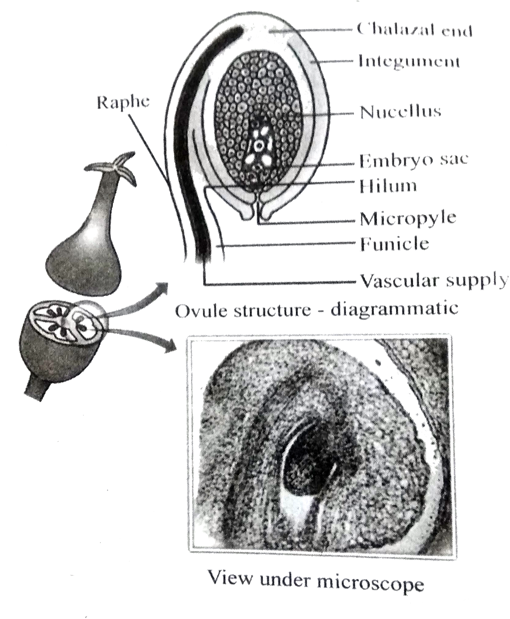
With a suitable diagram explain the structure of an ovule.Solution in Tamil
The ovule is otherwise called as megasporangium. It is covered by two integuments. Mature ovule has a stalk and a body. The stalk attaches the ovule to the placenta and is called funiculus or funicle. Hilum is the point at which the body of the ovule is attached to the funicle. Raphe is the ridge in an inverted ovule, in which the funicle is adnate to the body of the ovule. Nucellus, a central mass of parenchymatous tissue constitute the body of the ovule. They have reserve food material. The nucellus is covered by one or two integuments, which leaves a free portion at the top and forms a pore called micropyle. The ovule with one integuments is called as unitegmic, whereas the ovule with two basal region of body of the ovule, where the nucellus, the integument and the funicle meet or merge. The embryo sac like structure in the nucellus towards the micropylar end.

The functional megaspore formed from the nucellus further develops into embryo sac. In some species, the inner layer of integument forms specialized cells to perform the nutritive function for the embryo sac and such tissue is called endothelium or integumentary tapetum. (For example : Asteraceae). Based on the p ostion of the sporogenous cells, the ovules are divided into two types such as tenuinucellate type, the ovule is hypodermal with a single layer of nucellar tissue around it. Whereas in crassinucellate type, the ovule is with sub hypodermal sporogenous cell. Normally tenuinucellate ovules. have very small nucellus, while crassinucellate ovules have fairly large nucellus. Hypostase is the group of cells found at the base of the ovule between embryo sac and chalaza and epistase is the thick walled cells found above the micropylar end.
Topper's Solved these Questions
- BOOK - PREMIERS PUBLISHERSCHAPTER - ASEXUAL AND SEXUAL REPRODUCTION IN PLANTSEXERCISE - OTHER IMPORTANT QUESTIONS & ANSWERS (I. CHOOSE THE CORRECT ANSWERS:)30videos
ASEXUAL AND SEXUAL REPRODUCTION IN PLANTS
- BOOK - PREMIERS PUBLISHERSCHAPTER - ASEXUAL AND SEXUAL REPRODUCTION IN PLANTSEXERCISE - OTHER IMPORTANT QUESTIONS & ANSWERS (II. ANSWER THE FOLLOWING:)15videos
ASEXUAL AND SEXUAL REPRODUCTION IN PLANTS
- BOOK - PREMIERS PUBLISHERSCHAPTER - BIODIVERSITY AND ITS CONSERVATIONEXERCISE - OTHER IMPORTANT QUESTIONS & ANSWERS (Answer the followlng)22videos
BIODIVERSITY AND ITS CONSERVATION
Similar Questions
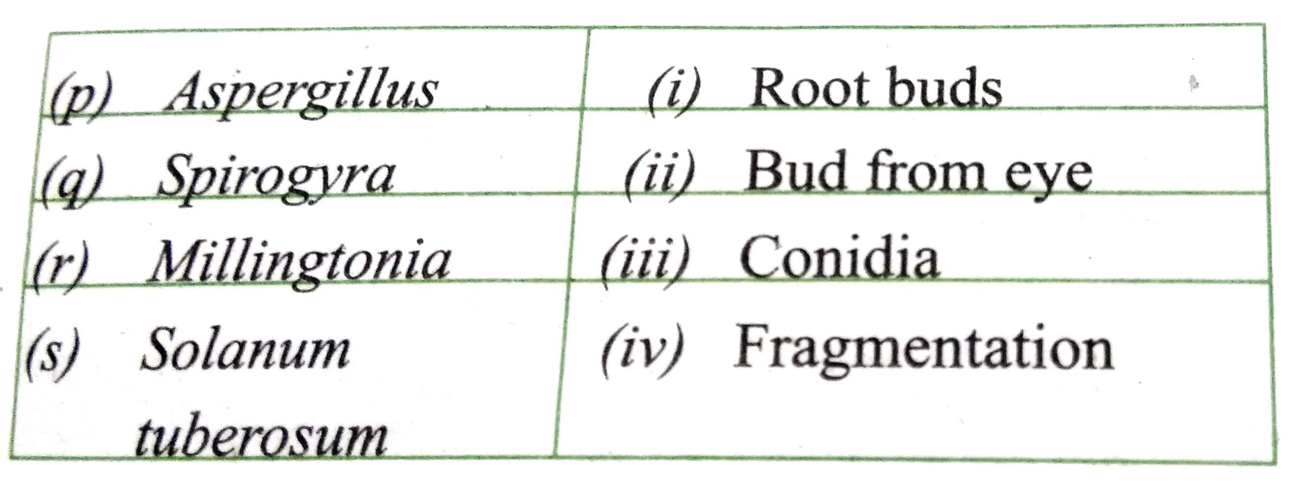
Match the following :
 02:50View Solution
Asexual reproduction
02:50View SolutionDefine biodiversity.
02:50View SolutionWith the help of suitable diagrams, explain how the age structures determine the trend of population ?
05:50View SolutionWith the help of suitable diagrams, explain how the age structures determine the trend of population ?
05:50View SolutionWith the help of suitable diagrams, explain how the age structures determine the trend of population ?
05:55View SolutionExplain the structure of immunoglobulin with suitable diagram.
04:04View SolutionExplain the structure of immunoglobulin with suitable diagram.
04:04View SolutionExplain the structure of chromosomes with suitable diagram.
03:24View SolutionWith a suitable diagram explain the structure of an ovule.
02:54View SolutionWith a suitable diagram explain the structure of an ovule.
05:31View SolutionDescribe the structure of anatropous ovule with labelled diagram.
03:45View Solutionफेफड़ों की रचना गैसीय आदान-प्रदान के लिए उपयुक्त होती है। चित्र द्वारा समझाइए।
03:18View SolutionWith a suitable diagram, explain the structure of an ovule.
05:50View SolutionExplain the structure of Immunoglobulin molecule with a suitable diagram.
02:05View SolutionWith a suitable diagram explain the structure of an ovule . Structure of ovule (Megasporangium) :
06:49View Solution
