Describe the T.S of a Monocot stem . Solution in Telugu
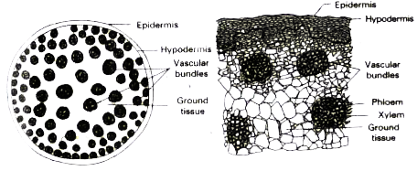
The structure of monocot stem can be understood well by observing the T.S of stem of Zea mays .It shows 4 distinct parts ., namely 1 . Epidermis , 2. Hypodermis 3. Ground tissue and 4 . Vascular bundles .
(1) Epidermis : It is the outermost layer composed of rectangular or tubular living cells arranged closely and compactly without intercellular spaces . The cells contain vacuolated protoplasts with a single nucleus but chloroplasts are absent . A waxy layer called .cuticle. is deposited on the external surface of the epidermis . Cuticle prevents the evaporation of water from the plant body . Trichomes are absent Numerous stomata are found in the epidermis through which exchange of gases occurs .
Epidermis gives protection to the inner tissues , helps in the exchange of gases and also prevents the evaporation of water .
Hypodermis : A distinct cortex is absent in Monocot stems . However a thick walled hypodermis is found beneath the epidermis .The cells of hypodermis are sclerenchymatous and are arranged compactly in 3-4 rows, without any intercellular spaces. It gives machanical strength of the stem .
Ground tissue : A major part of the stem is formed by an extensive soft , parencymatous tissue called the ground tissue . The peripheral layer consists of smaller cells while the inner layers show bigger cells . The cells of peripheral layers are chlorenchymatous and are connected with a assimilation of food materials .the inner layers of cells are concerned with storage of food materials . In monocot stem endodermis is absent .
Vascular bundles : Numerous vascular bundles are found scattered irregularly in the ground tissue . This kind of arrangement is called .atactostele. .It is considered an on advanced character . The inner vascular bundles are bigger in size and far apart from one another . The outer vascular bundles are smaller and are close to one another and found in one or teo circles .
Each vascular bundle is oval in shape and shows xylem and phloem together on the same radius . There is no cambium between xylem and phloem at the upper side of the vascular bundle . Each vascular bundle is enchosed by a sheath of sclenrenchymatous fibres . Hence it is called fibro vascular bundle .
Xylem consists of tracids , vessels ,fibres and parenchyma . Xylem vessels are few in number (4) and are arranged in "Y" shape . one or two protoxylem cells are crushed forming lysigeneous cavity called protoxylem lacunae which store water pholem consists of sieve tubes and companion cells . Phloem parenchyma is absent Medulla , Medullary rays and pericycle are also absent .

Related Playlists
- BOOK - VIKRAM PUBLICATION ( ANDHRA PUBLICATION)CHAPTER - HISTOLOGY AND ANATOMY OF FLOWERING PLANTS EXERCISE - TEXTUAL EXERCISES3videos
HISTOLOGY AND ANATOMY OF FLOWERING PLANTS
- BOOK - VIKRAM PUBLICATION ( ANDHRA PUBLICATION)CHAPTER - HISTOLOGY AND ANATOMY OF FLOWERING PLANTS EXERCISE - IMPORTANT QUESTIONS7videos
HISTOLOGY AND ANATOMY OF FLOWERING PLANTS
- BOOK - VIKRAM PUBLICATION ( ANDHRA PUBLICATION)CHAPTER - HISTOLOGY AND ANATOMY OF FLOWERING PLANTS EXERCISE - SHORT ANSWER TYPE QUESTIONS10videos
HISTOLOGY AND ANATOMY OF FLOWERING PLANTS
- BOOK - VIKRAM PUBLICATION ( ANDHRA PUBLICATION)CHAPTER - ECOLOGY AND ENVIRONMENT EXERCISE - IMPORTANT QUESTIONS45videos
ECOLOGY AND ENVIRONMENT
- BOOK - VIKRAM PUBLICATION ( ANDHRA PUBLICATION)CHAPTER - LOCOMOTION AND REPRODUCTION IN PROTOZOAEXERCISE - Important Questions13videos
LOCOMOTION AND REPRODUCTION IN PROTOZOA
Similar Questions
Name the various kinds of cell layers which constitute the bark .
04:36View SolutionWhy are xylem and phloem called complex tissues?
02:25View SolutionState the location and function of different types of meristem.
View SolutionDefine the term "ecology" and its branches.
05:58View SolutionList any two differences between a flagellum and a cilium.
03:31View SolutionDescribe the vascular bundles of monocot stem.
05:59View SolutionDescribe the internal structure of a monocot stem.
07:24View SolutionCompare T.S. of monocot and dicot stem with the help of well labelled diagrams only.
18:11View SolutionDescribe internal structure of monocot stem.
07:17View SolutionDescribe the T.S. of dicot stem.
02:50View SolutionWith the help of a neat labelled diagram, describe the T.S. of a monocot root.
01:52View SolutionWith the help of a neat labelled diagram, describe T.S. of dicot stem (sunflower).
View SolutionDraw and label a part of T.S. of a monocot stem. Explain various tissues present in it.
View SolutionDescribe the T.S of a dicot stem .
View SolutionDescribe the anatomy of monocot stem.
View Solution


