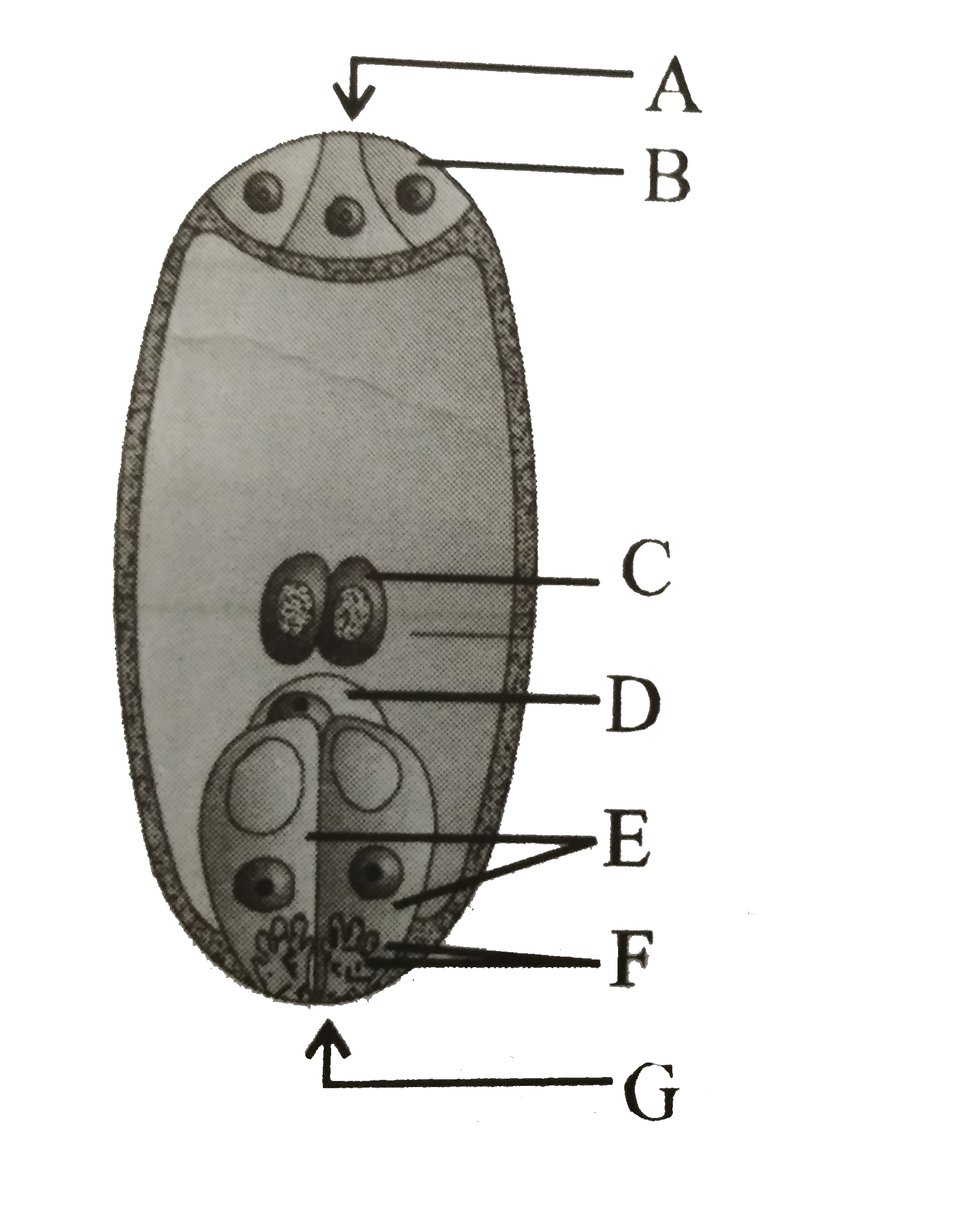
The given figure is associated to double fertilization A, B, C, D, E, F and G are identified respectively
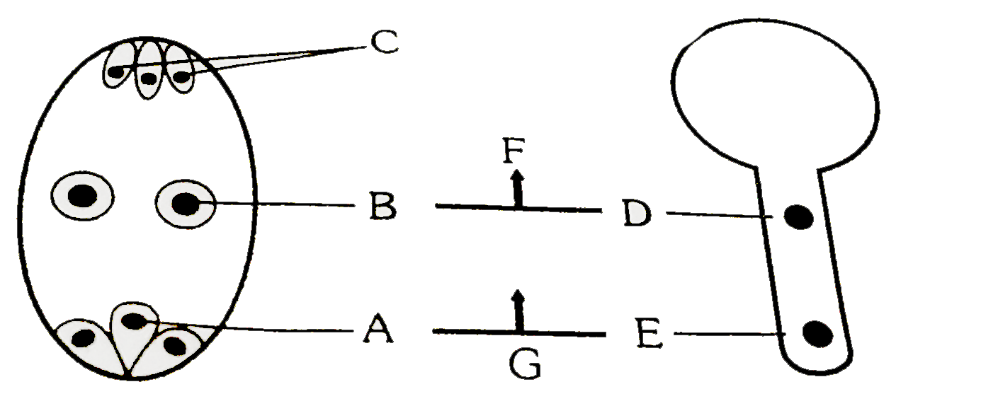
Egg, Polar nuclei, Male gamete, Male gamete, Zygote and Primary endosperm nucleus (PEN)
Egg, Male gamete, Male gamete, Polar nuclei, Primary endosperm nucleus (PEN) and Zygote
Egg, Male gamete, Polar nuclei, Male gamete, Primary endosperm nucleus (PEN) and Zygote
Egg, Polar nuclei, Male gamete, Male gamete, Primary endosperm nucleus (PEN) and Zygote
The correct Answer is:D
Step by step video & text solution for The given figure is associated to double fertilization A, B, C, D, E, F and G are identified respectively by Biology experts to help you in doubts & scoring excellent marks in Class 12 exams.
Updated on:21/07/2023Similar Questions
Knowledge Check
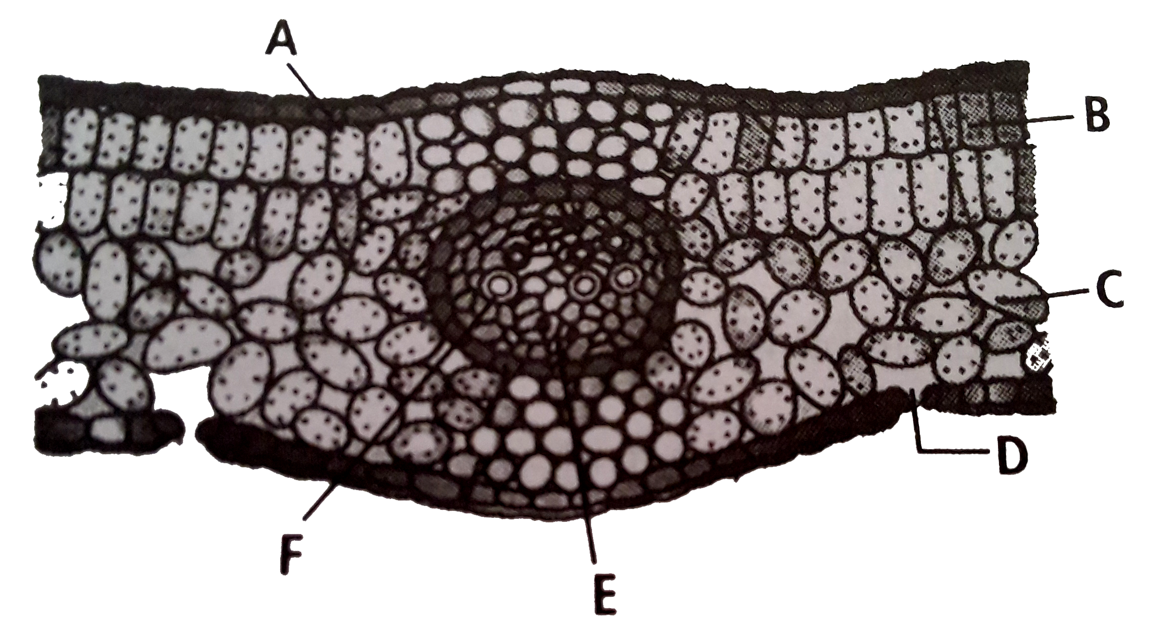
The given figure shows T.S of helianthus leaf with various parts labelled as A, B, C, D, E, F and G. identify the parts and select the correct optionAA-Epidermic, B-Spongy parenchyma, C-Palisade parenchyma, D-stomata, E-Phloem, F-XylemBA-Epidermis, B-Palisade parenchyma, C-Spongy parenchyma, D-Stomata, E-Xylem, F-PhloemCA-Epidermis, B-Palisade parenchyma, C-spongy parenchyma, D-Stomata, E-Endodermis, F-XylemDA-Epidermis, B-Palisade parenchyma, C-Spongy paranchyma, D-stomata, E-Phloem, F-XylemThe given figure shows T.S. of Mango leaf with various parts labelled as A, B, C, D, E, F and G. Identify the parts and select the correct option.
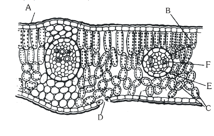 .AA-Epidermis, B-Spongy parenchyma, C-Palisade parenchyma, D-Stomata, E-Phloem, F-Xylem.BA-Epidermis, B-Palisade parenchyma, C-Spongy parenchyma, D-Stomata, E-Xylem, F-Phloem.CA-Epidermis, B-Palisade parenchyma, C-Spongy parenchyma, D-Stomata, E-Bundle sheath, F-Xylem.DA-Epidermis, B-Palisade parenchyma, C-Spongy parenchyma, D-Stomata, E-Phloem, F-Xylem
The given figure indicates the development of root nodule in soyabean
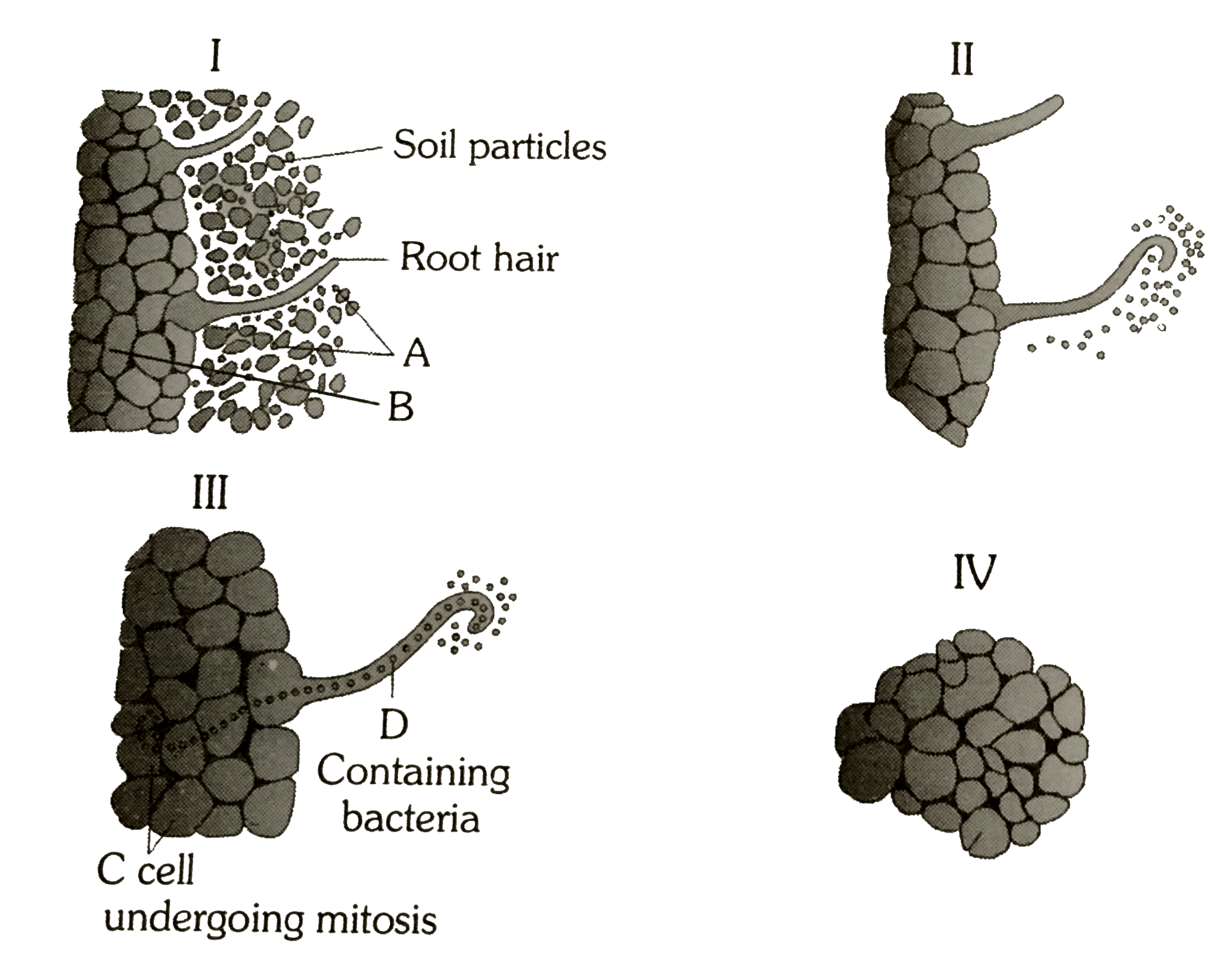
Identify A, B, C and D respectivelyAA- Nitrosomonas bacteria, B- Cortex cell, C - Inner cortex, D - Infection threadBA - Rhizobial bacteria, B - Endodermal cell , C - Inner Endodermis, D- Infection threadCA - Rhizobial bacteria, B - Cortex cell, C- Inner cortex, D - Infection threadDA - Rhizobial bacteria, B - Cortex cell, C - Outer cortex, D - Infection thread
The given figure show the structure of neuron,Identify A,B,C,D and E
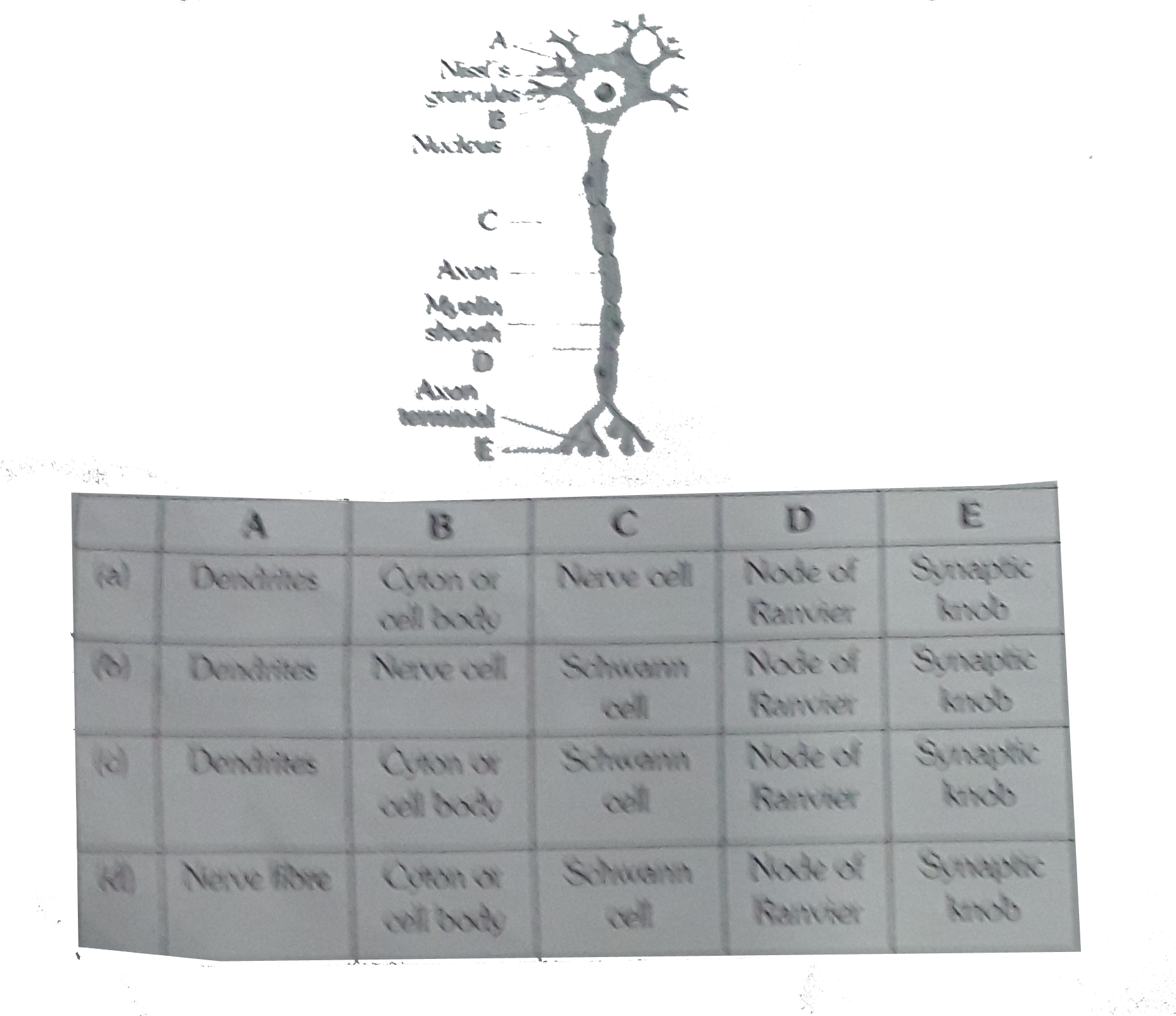
View SolutionIdentify A, B, C, D, E and F in the table given below :

View SolutionIdentify A, B, C, D, E in given diagram
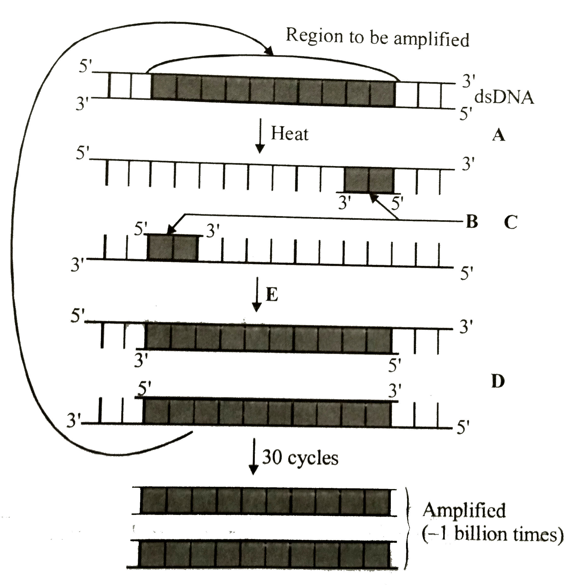
View SolutionThe given figure is associated with myosin monomer (meromyosin). Identify A to C
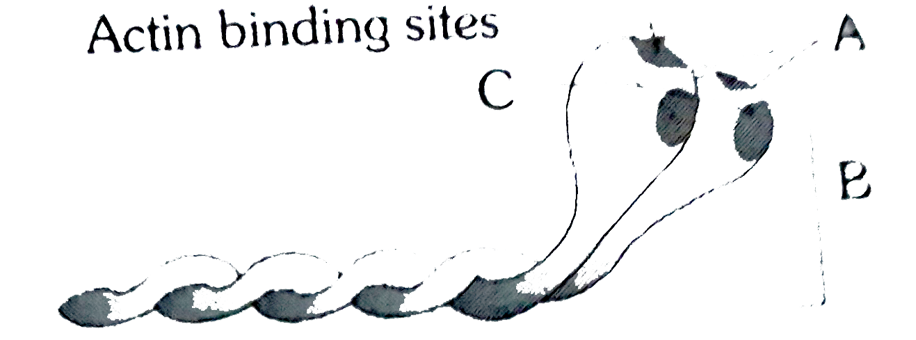
View SolutionSee the following figure and identify A, B, C, D and E are respectively
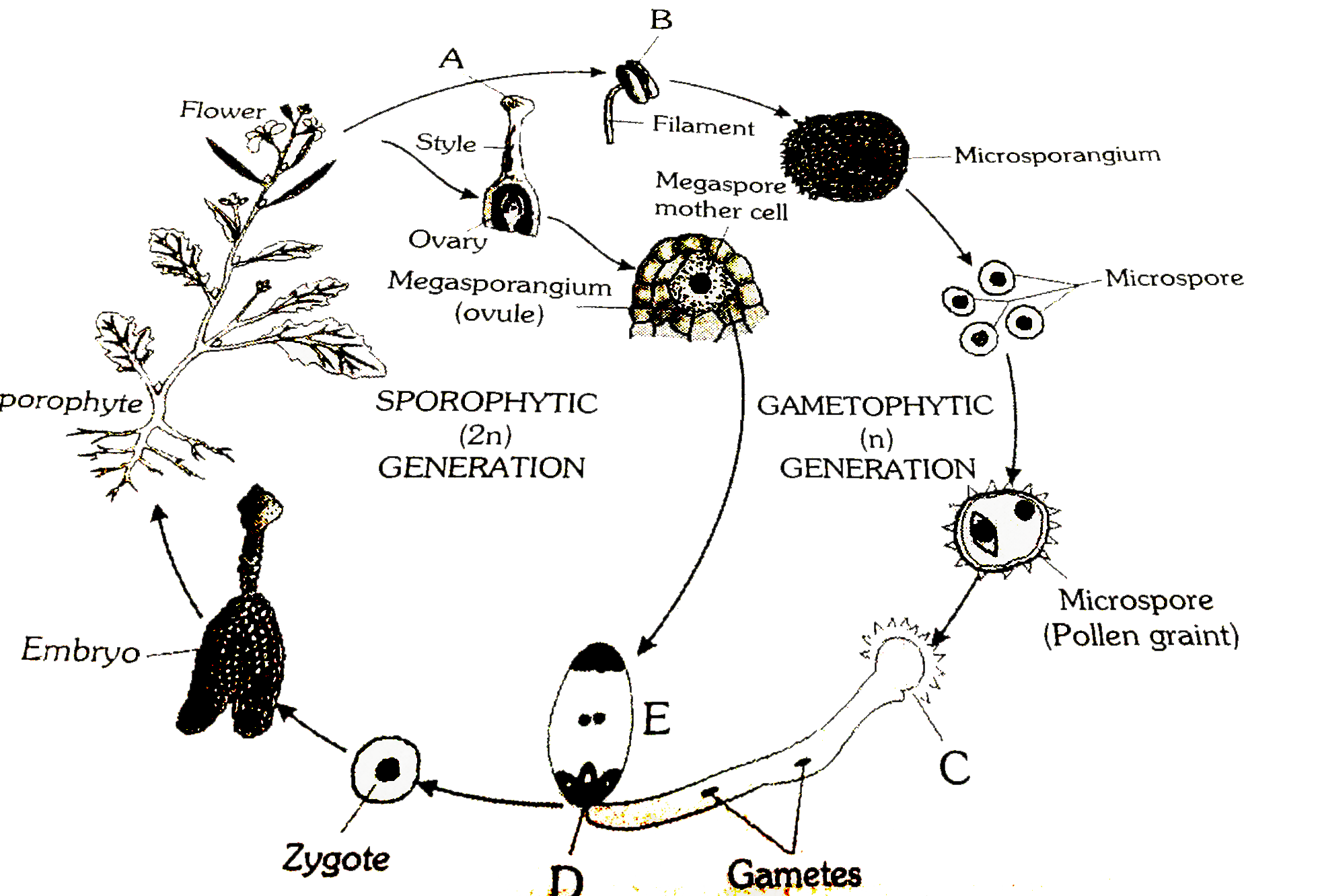
View Solution

