The given figure indicates the development of root nodule in soyabean
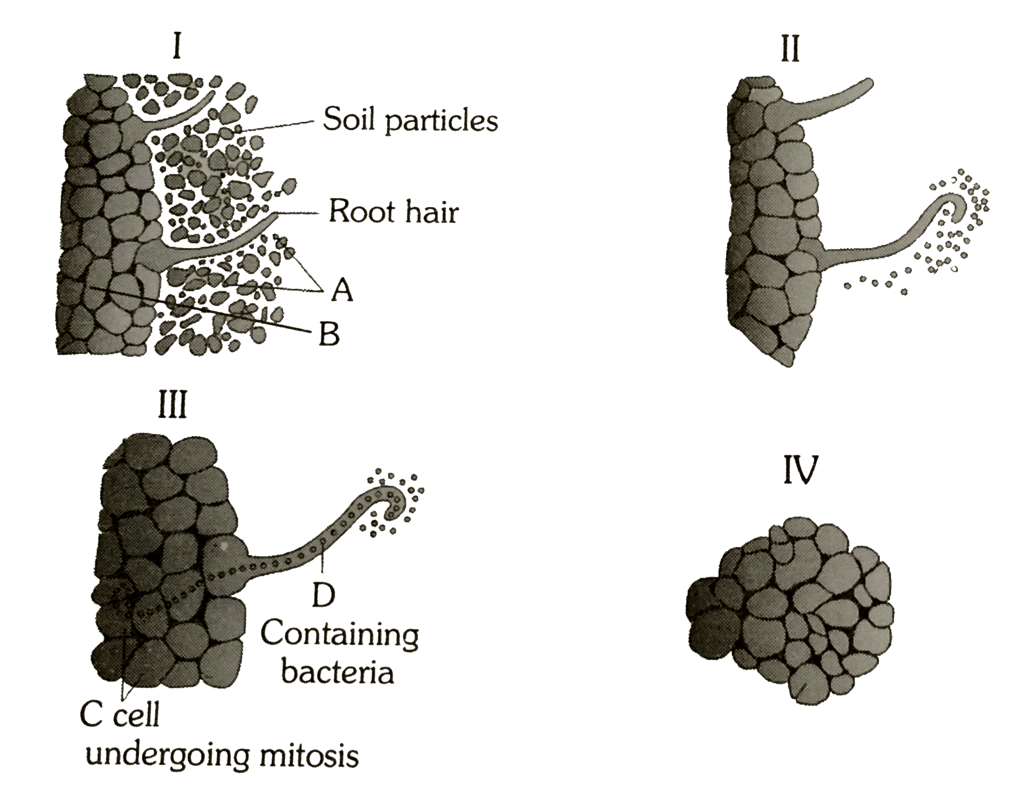
Identify A, B, C and D respectively
A - Nitrosomonas bacteria, B - Cortex cell, C - Inner cortex, D - Infection thread
A - Rhizobial bacteria, B - Endodermal cell, C - Inner Endodermis, D - Infection thread
A - Rhizobial bacteria, B - Cortex cell, C - Inner cortex, D - Infection thread
A - Rhizobial bacteria, B - Cortex cell, C - Outer cortex, D - Infection thread
The correct Answer is:C
Rhizobium multiplies and colonize the surroundings of roots and gets attached to epidermal and roots hair cells. This occurs because legume roots secrete some specific chemicals which attract the bacteria.
* Bacteria collect over the root hairs release Nod factors that cause curling of root hairs and the bacteria invades them. An infection thread containing the bacteria is produced. Infection thread grows along with multiplication of bacteria. * Infected thread carries the bacteria to the inner cortex where they initiate the nodule formation. * The invading bacteria stop dividing and get modified into rod-shaped structure called bacteroids. * Also the bacteria release signals that cause inner cortical and pericycle cell to divide. * Division and growth of cortical and pericycle cells lead to nodule formation.
Similar Questions
Identify the A,B,C and D are, respectively .

View SolutionStudy the given figure which shows the development of root nodules in soyabean. Match the correct explanation and find the correct sequence for labelled diagrams.
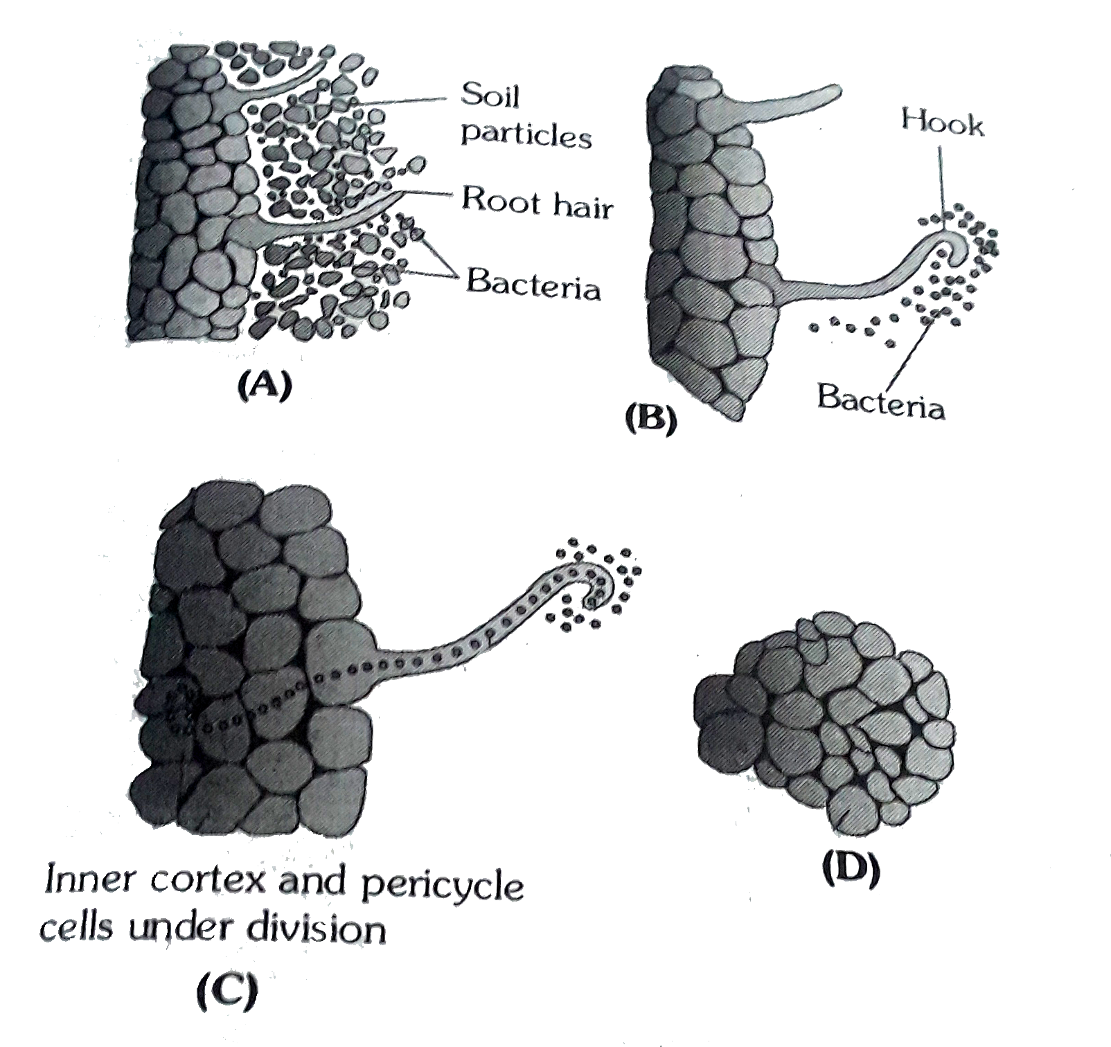
(I) Rhizobium bacteria contacting a susceptible root hair.
(II) A mature nodule having vascular connection with roots.
(III). Infection thread carrying bacteria to the inner cortex.
(IV). Curling of root hair due to successful infection.
View Solution
Knowledge Check
The following diagram indicates the development of root nodule in soyabean.

Identify A to D respectively.AA-Rhizobial bacteria, B - Cortex cell, C- Outer cortex, D-Infection threadBA-Rhizobial bacteria, B- Cortex cell, C - Inner cortex, D-Infection threadCA-Rhizobial bacteria, B- Endodermal cell, C- Inner Endodermis, D - Infection threadDA-Nitrosomonas bacteria, B-Cortex cell, C- Inner cortex, D-Infection threadIdentify A, B, C and D in given figure
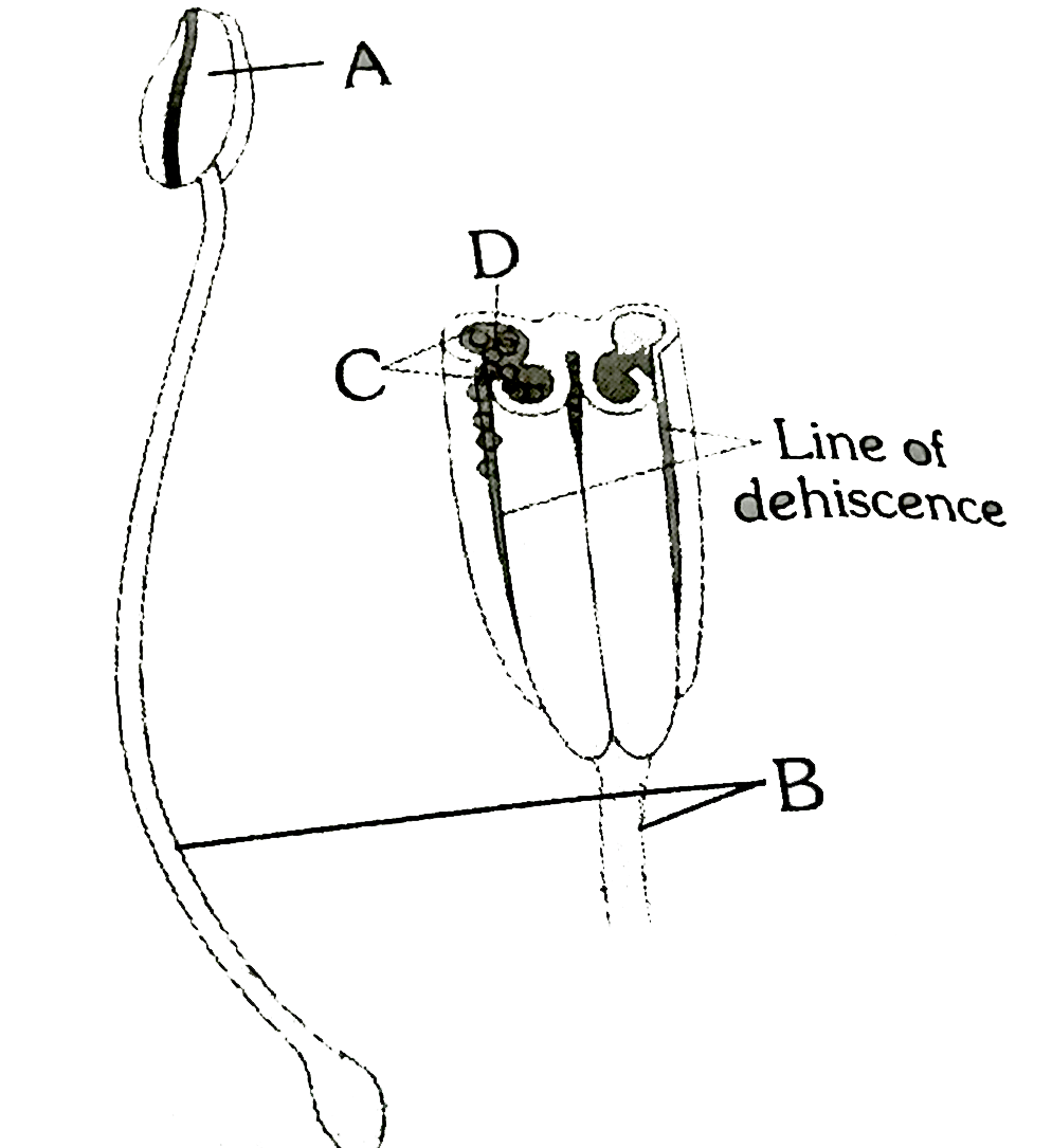 AA B C DAnther Filament Pollen sac Pollen grainsBA B C DAnther Pedicel Megasporangium Pollen grainsCA B C DAnther Petiole Megasporangium Pollen grainsDA B C DAnther Petiole Pollen sac Magaspore
See the following figure and identify A, B, C, D and E are respectively
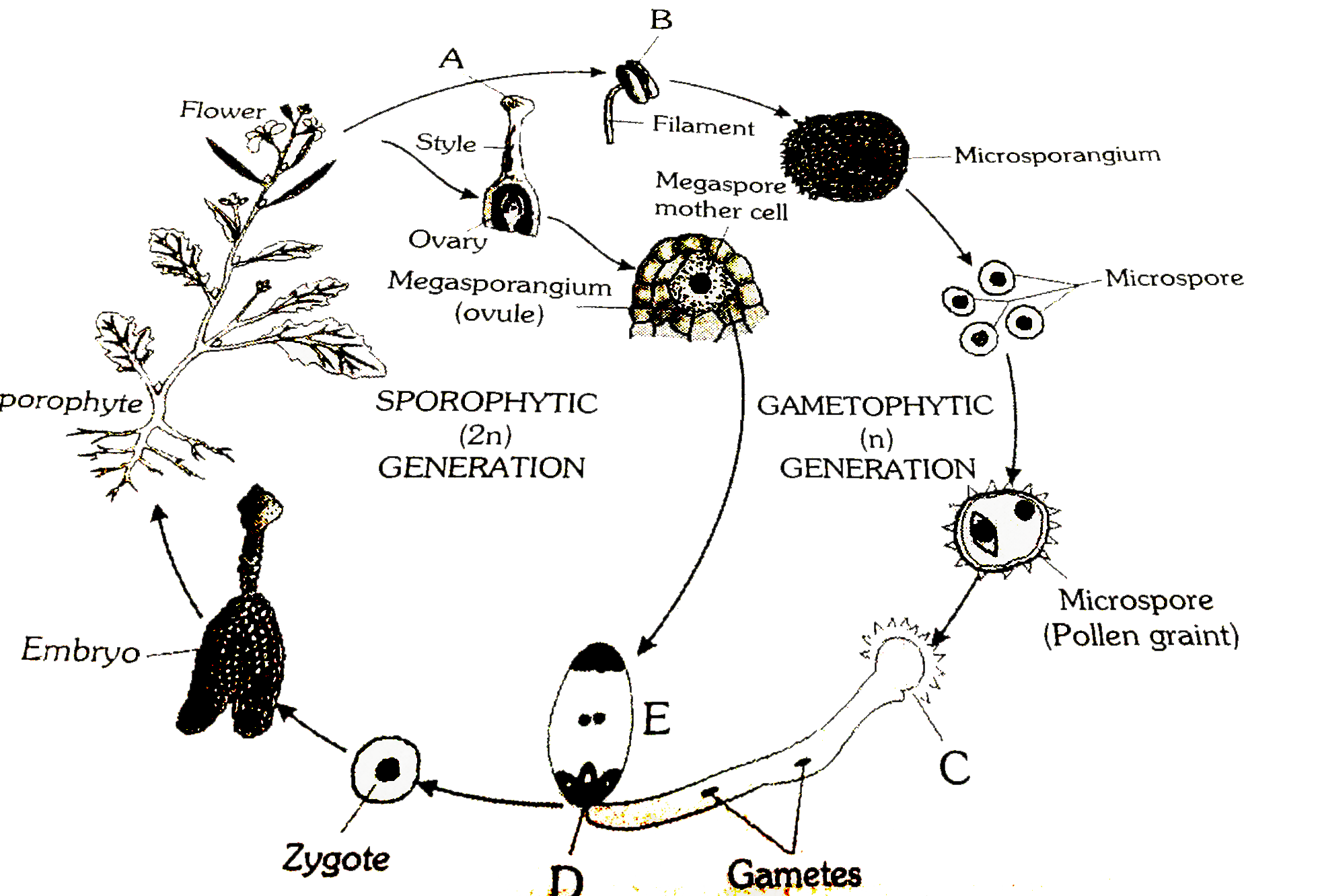 AStigma, Anther, Ebryo sac, Egg and female gametophteBStigma, Anther, Male, gametophyte, fertilized egg and female gametophyteCStigma, Anther, Female gametophyte Egg and Male gametophyteDStigma, Anther, Male gametophyte, Egg and female gametophyte
In the given figure , identify A, B , C , D and E respectively.

View SolutionIdentify A,B,C and D in the given table:

View Solution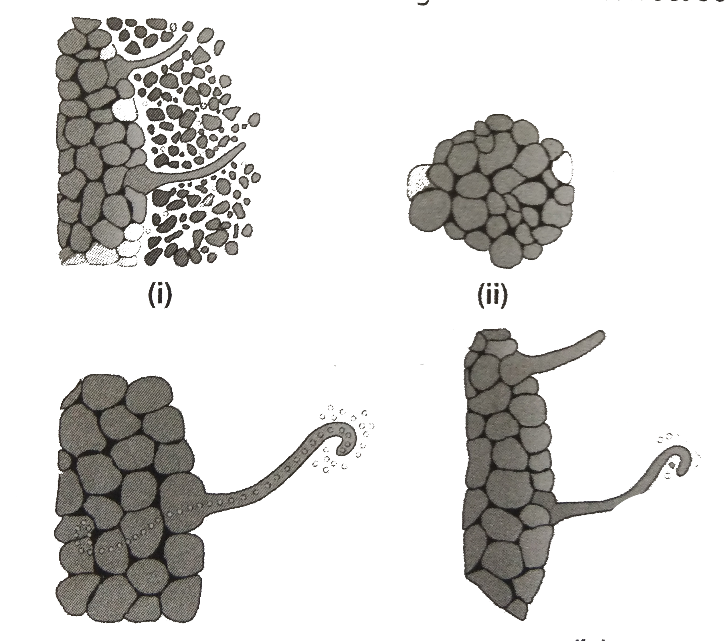
Study the given figures carefully showing the development of root nodules and arrange them in a correct sequence.
View SolutionThe given figure indicates the stages of seed gremination
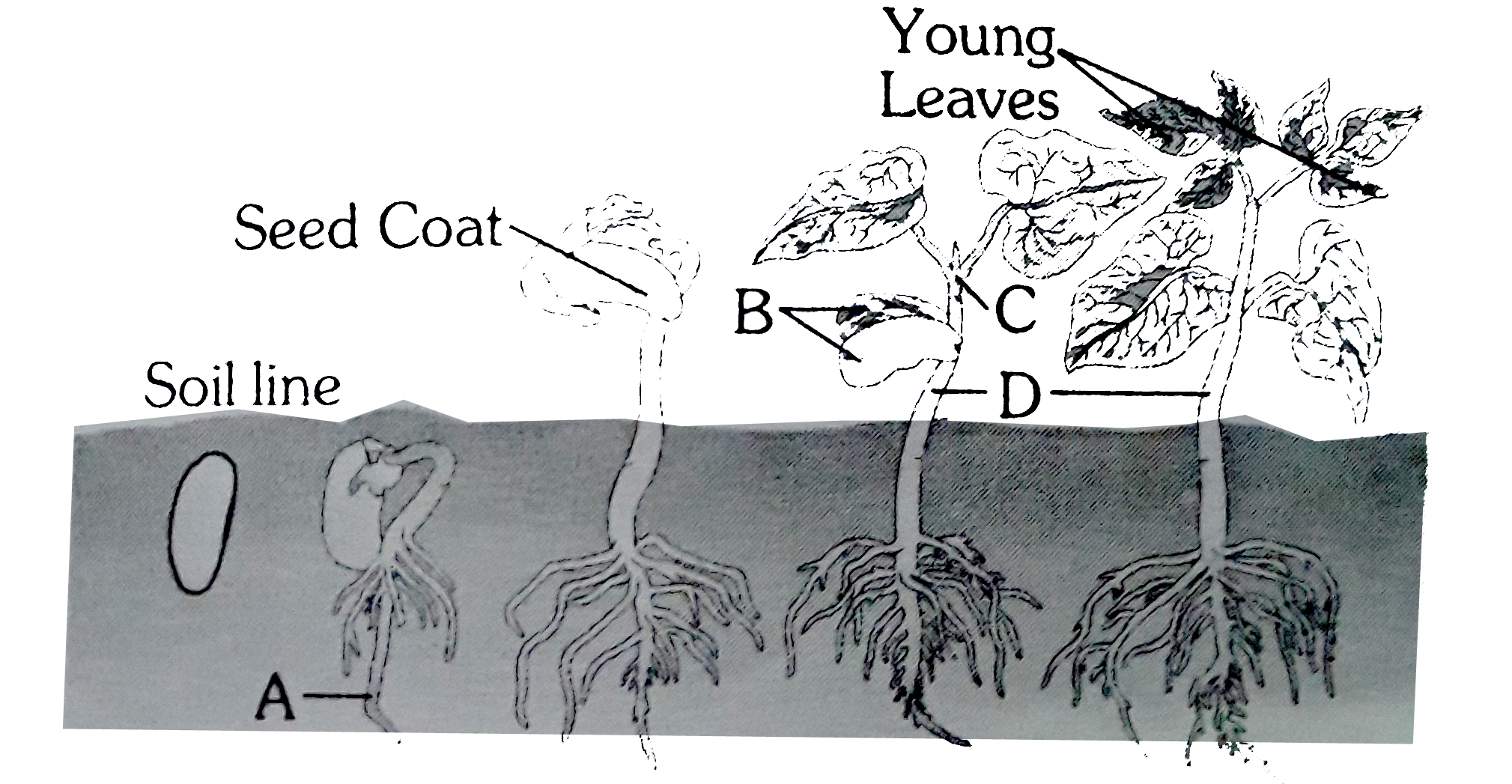
Identify A,B,C and D respectivelty
View SolutionSee the figure given below and identify A to D respectively

View Solution
