Draw the structure of lungs and label the fol- lowing parts in it.
(1) Nasal cavity (2) Epiglottis (3) Lungs (4) Larynx and vocal cords (5) Trachea (6) Diaphragm (7) Bronchus (8) Alveoli
To draw the structure of the lungs and label the specified parts, follow these steps:
1. Draw the Basic Outline of the Lungs:
- Start by sketching two large, oval shapes to represent the left and right lungs. The right lung is typically larger and has three lobes, while the left lung has two lobes due to the space occupied by the heart.
2. Add the Trachea:
- Draw a vertical tube extending downwards from the throat area to represent the trachea (windpipe). This should be positioned centrally above the lungs.
3. Include the Bronchi:
- At the bottom of the trachea, draw two branches extending from it, one leading into each lung. These are the bronchi (singular: bronchus).
4. Draw the Alveoli:
- At the end of the bronchi, draw small clusters of tiny sacs to represent the alveoli. These should look like small grapes or balloons and are located at the ends of the bronchi.
5. Add the Larynx:
- Above the trachea, draw a small box-like structure to represent the larynx. This is located at the top of the trachea.
6. Include the Vocal Cords:
- Inside the larynx, you can draw two thin lines to represent the vocal cords.
7. Draw the Nasal Cavity:
- Above the larynx, sketch a cavity that represents the nasal cavity. This should be a larger space above the larynx and trachea.
8. Add the Epiglottis:
- Draw a flap-like structure at the base of the larynx that covers the trachea when swallowing. This is the epiglottis.
9. Draw the Diaphragm:
- Below the lungs, draw a dome-shaped structure to represent the diaphragm, which separates the thoracic cavity from the abdominal cavity.
10. Label All Parts:
- Clearly label each part: Nasal cavity, Epiglottis, Lungs, Larynx and vocal cords, Trachea, Diaphragm, Bronchus, and Alveoli.
- Start by sketching two large, oval shapes to represent the left and right lungs. The right lung is typically larger and has three lobes, while the left lung has two lobes due to the space occupied by the heart.
- Draw a vertical tube extending downwards from the throat area to represent the trachea (windpipe). This should be positioned centrally above the lungs.
- At the bottom of the trachea, draw two branches extending from it, one leading into each lung. These are the bronchi (singular: bronchus).
- At the end of the bronchi, draw small clusters of tiny sacs to represent the alveoli. These should look like small grapes or balloons and are located at the ends of the bronchi.
- Above the trachea, draw a small box-like structure to represent the larynx. This is located at the top of the trachea.
- Inside the larynx, you can draw two thin lines to represent the vocal cords.
- Above the larynx, sketch a cavity that represents the nasal cavity. This should be a larger space above the larynx and trachea.
- Draw a flap-like structure at the base of the larynx that covers the trachea when swallowing. This is the epiglottis.
- Below the lungs, draw a dome-shaped structure to represent the diaphragm, which separates the thoracic cavity from the abdominal cavity.
- Clearly label each part: Nasal cavity, Epiglottis, Lungs, Larynx and vocal cords, Trachea, Diaphragm, Bronchus, and Alveoli.
Topper's Solved these Questions
- LIFE PROCESSES IN HUMAN BEINGS - II (RESPIRATORY AND CIRULATORY SYSTEMS)BOOK - PEARSON IIT JEE FOUNDATIONCHAPTER - LIFE PROCESSES IN HUMAN BEINGS - II (RESPIRATORY AND CIRULATORY SYSTEMS)EXERCISE - MASTERING THE CONCEPTS (Application and Analysis)
20 Videos
- LIFE PROCESSES IN HUMAN BEINGS - II (RESPIRATORY AND CIRULATORY SYSTEMS)BOOK - PEARSON IIT JEE FOUNDATIONCHAPTER - LIFE PROCESSES IN HUMAN BEINGS - II (RESPIRATORY AND CIRULATORY SYSTEMS)EXERCISE - MASTERING THE CONCEPTS (Assertions and Reasons)
9 Videos
- LIFE PROCESSES IN HUMAN BEINGS - II (RESPIRATORY AND CIRULATORY SYSTEMS)BOOK - PEARSON IIT JEE FOUNDATIONCHAPTER - LIFE PROCESSES IN HUMAN BEINGS - II (RESPIRATORY AND CIRULATORY SYSTEMS)EXERCISE - QUICK RECAP
5 Videos
- LIFE PROCESSES IN HUMAN BEING-I (DIGESTIVE AND EXCRETORY SYSTEMS)BOOK - PEARSON IIT JEE FOUNDATIONCHAPTER - LIFE PROCESSES IN HUMAN BEING-I (DIGESTIVE AND EXCRETORY SYSTEMS)EXERCISE - ASSESSMENT TESTS (ANSWER THE QUESTIONS)
7 Videos
- LIFE PROCESSES IN PLANTSBOOK - PEARSON IIT JEE FOUNDATIONCHAPTER - LIFE PROCESSES IN PLANTSEXERCISE - ASSESSMENT TESTS (ANSWER THE FOLLOWING QUESTIONS)
14 Videos
Similar Questions
Draw the human respiratory system and label the following parts:
(a) Trachea
(b) Alveoli
(C ) Respiratory bronchioles
(d) Larynx.
Describe in brief the role of lungs in the exchange of gases.
View Solution1.Draw number lines and locate the points on them: (a) 12,14,34,44 (b) 18,28,38,78 (c) 25,35,85,45
View Solution
Knowledge Check
Read the following statements
(i) the point of bifurcation of trachea is called carina and is at the level of 5th thoracic vertiebra
(ii)the right bronchus is shorter iwder and more in line with trachea than the let bronchus
(iii) the bronchioles are without cartilaginous rings
(iv) the surfactant of lungs is secrated in infants between 6th and 7th month o flife
which of these are correct ?AI,ii & iiiBii , iii & ivCI, iii & ivDall are correctFind out the correct match from the following :−
(i) Larynx -sound box
(ii) Trachea - complete cartilagenous rigns
(iii) Epiglottis -elastic cartilagenous flap
(iv) Alveoli - very thin, irregular walled, non-vascularised bag like structured
(v) Pleural fluid - reduces friction on the lung surfaceA(i),(ii),(iii),(iv),(v)B(i),(iii),(iv),(v)C(i),(iii),(v)D(i) and (v)Which of the following comprises the lungs?
a. Tracheae, b. 1∘bronchi, c. 2∘ bronchi, d. 3∘ bronchi, e. initial bronchioles, f. terminal bronchioles, g. duct of alveoli, h. alveoliAb, c, d, e, f, g and hBa,b,c,d,e,f and gCa, b,c,d,e and fDa,b,c,d and e

Find the value of Min[(1#2) $ 3, 4#(5$6)] $ Max (6#7), (7#8)] :
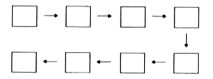
View SolutionS to Z are structures of the human respiratory system. Arrange them to show the order in which air from outside travels through the respiratory system to reach the gaseous exchange surface in the lungs.
S: Bronchioles
U: Nostrils
W: Epiglottis
Y: Trachea
T: Bronchi
V: Pharynx
X: Larynx
Z: Alveoli

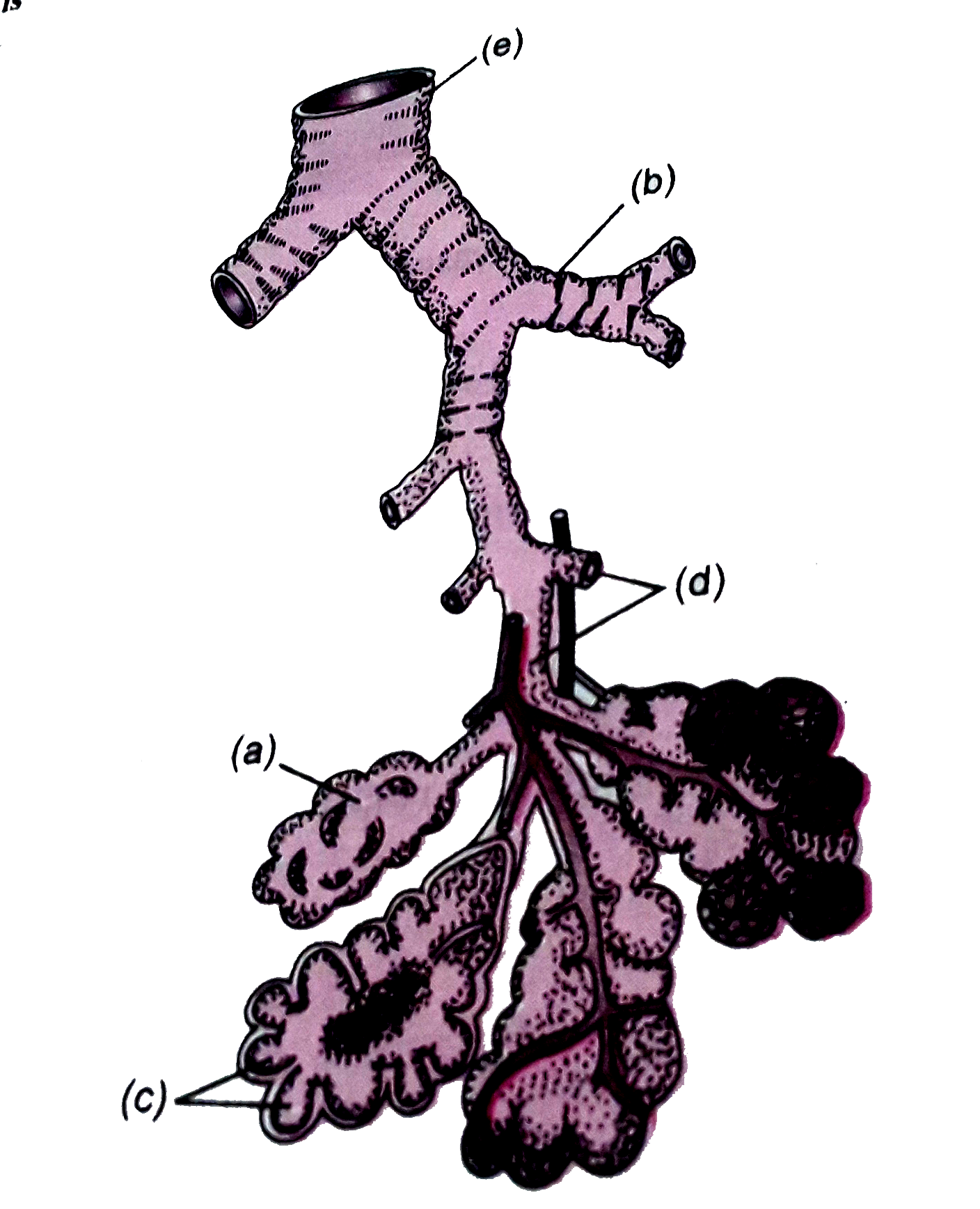
View Solution(i) Study the given figure of respiratory passage carefully . Parts are labelled as a,b,c,d and e. Label these parts .
(ii) How many secondary bronchi are there in right and left human lung ?
(iii) What is the approximatge length width of human trachea (wind pipe) ?
(iv) What is the role of epiglottis?
(v) In which body cavity the lungs are located ?

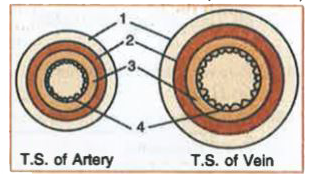
View SolutionIdentify structures 1,2,3 ,4 from the given diagram.

(b) Fill in the blanks . The poorly oxygenated blood comes from the body parts and poured into (1) and then pumped into (2) from which, through (3) artery to lungs. Then from lungs oxygenated blood enters into (4) through pulmonary (5). Then pumped into (6) from this the oxygenated blood passes through (7) to the body except lungs.
View SolutionWhat should come next in the following series?
9 8 7 6 5 4 3 2 1 9 8 7 6 5 4 3 2 9 8 7 6 5 4 3 9 8 7 6 5 4 9 8 7 ?
View Solution
