Describe the head of cockroach.
Text Solution
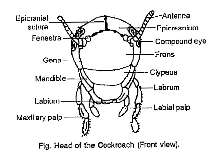
A. Head. It is the anterior most region of the body of a cockroach, which lies at right angle to the long axis. The proximal, semi-circular broader end of the head is-directed upwards, while the distal, narrow, mouth bearing end is directed downwards. This type of head is called hypognathus. The neck allows the movement of the head in all possible directions.

(i) Sclerites. The sclerites of the head are fused to form a head capsule. The top of the head capsule is known as vertex. The vertex is divided by epicranial suture into two epicranial plates. A triangular plate, called frons is found in front of the epicranial plates. The frons is followed by a rectangular plate, the clypeus. A frontoclypeal suture is present in between the frons and clypeus. The labrum, hangs from the lower edge of the clypeus. A labroclypeal suture is found in between the clypeus and labrum. Two cheek sclerites, the genae, lie vertically just below the eye on the lateral sides.
(ii) Sense organs. The sense organs situated on the head are :
a) compound eyes (b) antennae (c) fenestrae.
(a) Compound eyes. Two large black coloured, kidney shaped dorsolaterally compound eyes are placed on the top of the head. They are formed by a large number of ommatidia. These are externally represented by the same number of hexagonal areas. The compound eyes are the organs of sight.

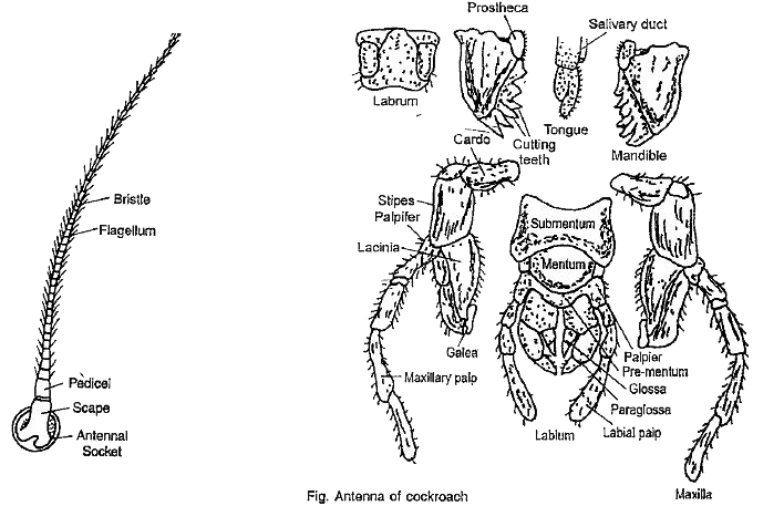
(b) Antennae. Just inner to the eye, a long, many jointed antenna arises from the antennal socket. It consists of three parts :
Scape. It is a large basal part of the antenna, which articulates with antennal socket.
Pedicel. It is a short and mid portion of the antenna.
Flagellum. The pedicel is followed by a long filamentous, many joined flagellum, which tapers distally.
The antennae can be moved in all directions. They possess sensory bristles which are tactile in nature. With the help of antennae, the cockroaches detect the presence of food.
(c) Fenestrae. A small rounded white area, the fenestra or ocellar spot is situated just towards .the inner and upper side of antennal socket. They represent the underdeveloped simple eyes.
(iii) Mouth parts. The mouth lies at the lower edge of head, and the appendages associated with the mouth are called mouth parts. They are used in searching and taking the food. The biting and chewing type of mouth parts has a Iabrum, two mandibles, two maxillae-a labium and a hypopharynx.
(a) Labrum. It is a broad, chitinous flap, which hangs the distal end of the clypeus. It is also called as upper lip. The· epipharynx, is fused to the inner surface of the labrum. The epipharynx bears the organs of taste. TheJabrum holds the food particles during the feeding.
(b) Mandibles. These are two stout, highly chitinous structures, articulating with the sides of the head capsule, and lying .below the genae. Inner margin of each mandible.bears teeth, while on its upper inner edge, a soft lobe, the prostheca, is present. The mandibles are operated by the abductor and abductor muscles for grinding the food.
(c) First pair of Maxillae. ·They lie beneath the mandibles on either side of the head. Each maxilla consists of three main parts :
(i) Protopodite. This is a basal portion, which is made up of two parts-the proximal cardo and the distal stipes. Both the parts are bent at an angle to each other.
(ii) Endopodite. It arises from the inner side of the s.tipes and consists of two parts - an outer broad, hook-like galea and an fnner, hard, platelike lacinia. The latter tapers into two sharp claw like projections. Lacinia also bears numerous strong sensbry bristles at its inner surfaces.
(iii) Exopodite. It consists of a small basal sclerite, the palpifer attached to the outer surface of the stipes, and a five joined maxillary palp.
Functions.
I. With the help of a lacinia, a cockroach holds the food and gives it to the mandibles for mastication.
2. Maxillae are also used for cleaning the antennae and the first pair of legs.
3. The bristles of the maxillary palps are tactile in nature.
( d) Labium or Second pair of maxilla. It is a single structure, which represents the fused first pair of maxillae, lying behind the mouth and forms a sort oflower lip. It comprises of three main parts.
(i) Protopodite. It consists ofa proximallarge part, the sub-mentum, the middle small mentum and the distal pre-mentum which represents the fused cardos, while pre-mentum is perhaps-the fused portion of two stripes.
(ii)Endopodite. Partially fused endopodites are represented by the ligula, whose each half consists of an inner glossa and an outer paraglossa which correspond to the lacinia and gal ea of the first pair of maxilla.
(iii) Exopodite. It consists of two parts. The small sclerite the palpiger, arises from the prementum on each side and a three jointed labial palp bearing sensory bristles attached to the palpiger. It corresponds to the maxillary palp of the first maxilla.
Functions:
I. Glossae and paraglossae prevent the loss of food prarticles from mandibular action.
2. The labial palps are sensory in nature.
(e) Hypopharynx. It is a small conical fleshy structure hanging in between the two maxillae, in front of the labium. An efferent salivary duct carrying the saliva from the salivary glands opens near the base of the hypopharynx. Hypopharynx directs the saliva towards the food.
|
Updated on:21/07/2023
Similar Questions
Describe the leg of a cockroach.
View SolutionHead of cockroach is
View SolutionDescribe trachea of cockroach and mode of tracheal respiration.
View SolutionThe head of cockroach lacks
View SolutionThe head of cockroach lacks
View SolutionThe endoskeleton in the head of cockroach is known as
View SolutionThe ingrowth of exoskeleton in the head of cockroach is called
View SolutionThe ingrowth of exoskeleton in the head of cockroach is called
View SolutionWrite the names of appendages associated with the head of a cockroach.
View SolutionMark the incorrect statement regarding the head of the cockroach.
View Solution

