What do you understand by the development of an embryo. Support your answer with the help of suitable diagrams. Solution in Punjabi
Video Solution
Text Solution
Embryonic Development
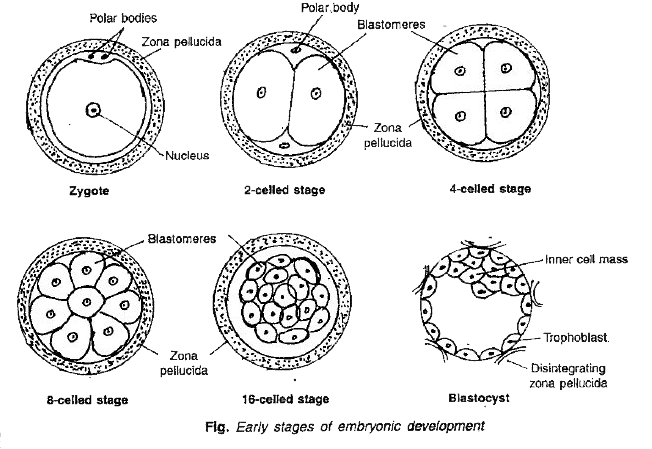
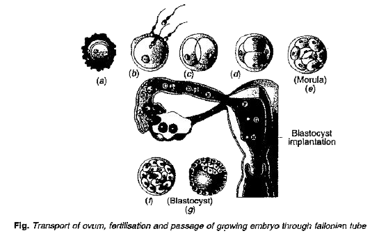
(i) About/thirty hours after fertilization, the zygote undergoes a series of mitotic divisions called cleavage and forms 2, 4, 8, 16 daughter cells called blastomeres. (ii) The embryo with 8 to 16 blastomeres and solid mass of cells is called morula (.Tittle mulberry.). The. morula continues to divide and transforms into blastocyst (Hollow ball of around 64 cells). The blastocyst remains free in the uterine cavity for several days before it implants in the uterine lining. The cavity of blastocyst is called blastocoel. (iii) The blastomeres in the blastocyst are arranged into an outer layer called trophoectoderm or trophoblast and an inner group of cells called the inner cell mass. (iv) The inner cell mass of the blastocyst is the precursor of the embryo i.e., forms the tissues of future embryo. The cells of trophoblast form the extraembryonic membranes and part of the placenta.
Implantation
(i) The attachment of the blastocyst to the uterine wall is called implantation, (ii) Implantation takes place about 7 days after fertilisation

(iii) The pole of the blastocyst that contains the inner cell mass attaches to the endometrium of the uterus. The trophoblast cells secrete proteolytic enzymes that erode the cells of the endometrium and blood vessels. It establishes the pregnancy. (iv) Zona pellucida layer plays a significant role in implantation. It prevent the implantation of the blastocyst at an abnormal site. When the blastocyst reaches the proper site, zone pellucida exposes the sticky phagocytic cells of trophoblast. Now zona pellucida disappears.
Formation of Placenta
(i) After implantation, finger like projections appear on the trophoblast called chorionic villi which are surrounded by the uterine tissue and maternal blood. The chorionic villi and uterine tissue become interdigitated with each other and jointly form a structural and functional unit between developing embryo and maternal body called placenta. Placenta is connected to embryo by umbilical cord. (ii) Placenta facilitates the supply of oxygen and nutrients from maternal blood to the embryo. The carbon-dioxide and waste materials produced by the embryo also pass to maternal blood through placenta and are excreted by the mother. (iii) Placenta also acts as an endocrine tissue and produces several hormones like human chorionic gonadotropin (HCG), human placental lactogen (HPL), estrogens, progestogens etc.
Formation of Germ layers (Gastrulation)
(i) The embryonic stage extends from the second through the eighth week of development. It starts with the completion of implantation and includes the formation of germ layers, embryonic membranes, and the placenta. The inner cell mass contains stem cells which have the potency to give rise to all tissues and organs. (ii) Embryonic development at various months of pregnancy : After one month, embryo.s heart is formed, end of second month limbs and digits are formed and by the end of third month limbs and external genital organs are formed. During 5th month movements of the foetus and appearance of hair on head are observed. In the end of second trimester body is covered with fine hair, eyelid separates and eyelashes are formed. By the end of 9th month, foetus is fully developed and ready for birth.

|
Updated on:21/07/2023
Similar Questions
विकास से आप क्या समझते हैं? (उत्तर 30 शब्दों में दीजिए)
View Solution(A) What do you understand by the molecularness of a reaction? Confirm your answer by giving suitable examples. (B) What do you understand by the order of the reaction? Give an example also.
04:51View SolutionWhat do you understand by heterotrophic mode of nutrition? Elaborate your answer with suitable examples.
04:05View Solutionविकास से आप क्या समझते हैं ?
View Solutionविकास से आप क्या समझते हैं ? ( उत्तर 30 शब्दों में दीजिए। )
View SolutionExplain the development of dicot embryo giving suitable example.
11:40View SolutionDescribe the development of monocot embryo with suitable diagrams.
06:59View SolutionWhat do you understand by multiplication choices? Explain with the help of suitable examples.
02:32View Solution
