Describe the structure of kidneys.Solution in Telugu
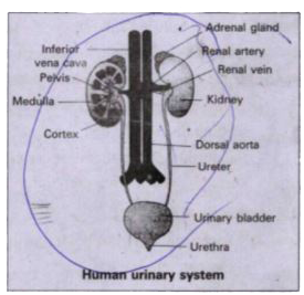
In humans, the excretory system consists of a pair of kidney, a pair of ureters, a urinary bladder and urethra.
Kidney : Kidneys are reddish brown, bean shaped structures, situated on either side of the vertebral column between the levels of last thoracic and third lumbar vertebrae in a retroperitoneal position. The right kidney is slightly lower than the left one due to the presence of liver.
The outer surface of the kidney is convex and the inner surface is concave, where it has a deep notch called hilum, the point at which the renal artery and nerves enter and renal vein and ureter leave. Each kidney is surrounded by a tough, fibrous tissue, called renal capsule.
Ureter : These are slender whitish tubes, which emerges from the pelvis of the kidney. The ureter rundown and open into the urinary bladder.
Urinary bladder : Urinary bladder is a pear shaped like muscular organ. It termpirarily stores the urine, situated in the lower abdominal cavity. The neck of the bladder leads into the urethra. Urethra opens near the vaginal orifice in the female and through the penis in the male.
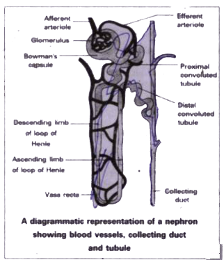
Structure of nephron : Each kidney has nearly one million nephrons. These are structural and functional units of kidney, embedded in the loose connective tissue of cortex and medulla. Nephron consist of malpighian body and renal tubule.

I) Malphigian body : It begins in the cortex of the kidney. It contains Bowman.s capsule and glomerulus.
Bowman.s capsule : It is a thin walled, double layered cup. The inner wall of the Bowman.s capsule has certain unique cells called podocytes.
Glomerulus : It is a dense network of capillaries in the cup of Bowman.s capsule. Afferent arteriole of renal artery enter the cavity of Bowman.s capsule and split into five branches. They unite and come out of the Bowman.s capsule as an afferent arteriole.
The podocytes of inner wall of Bowman.s capsule wrap around each capillary. The podocytes are arranged in an intricate manner so as to leave some minute spaces called filteration slits. The endothelium cells of capillaries have numerous pores called fenestrations.
II) Renal tubule : It is narrow, delicate tubule arises from the posterior part of Bowman.s capsule known as neck. It opens into along narrow convoluted tubule with three parts like proximal convoluted tubule, Loop of Henle and Distal convoluted tubule.
a) Proximal convoluted tubule : It is a lined by simple cuboidal eptihelium with brush border to increase area of absorption.
b) Loop of Henle : It is a hairpin like tubule present in medulla region. It consist of a descending limb and an ascending limb. The proximal part of the ascending limb is thin and the distal part is thick. The thick ascending limb continuous into the distal convoluted tubule.
c) Distal convoluted tubule (DCT) : It is present in cortex. It is lined by simple cuboidal epithelium. The DCT continuous as the initial collecting duct in the cortex.
Collecting system : Some initial collecting ducts unite to form straight collecting duct, which passes through the medullary pyramid. In the medulla, the tubes of each pyramid join and form duct of Bellini, which finally opens into the tip of the renal papilla.

Capillary network of nephron : The efferent arteriole emerging from the glomerulus forms a fine capillary network called the peritubular capillaries, around the renal tubule. The portion of the peritubular capillaries that surrounds the loop of Henle is called the vasa recta. The vasa recta is absent or highly reduced in the cortical nephrons. The juxta medullary nephrons posses well developed vasta recta.
Related Playlists
- BOOK - VIKRAM PUBLICATION ( ANDHRA PUBLICATION)CHAPTER - EXCRETORY PRODUCTS AND THEIR ELIMINATIONEXERCISE - SHORT ANSWER QUESTIONS13videos
EXCRETORY PRODUCTS AND THEIR ELIMINATION
- BOOK - VIKRAM PUBLICATION ( ANDHRA PUBLICATION)CHAPTER - ENZYMESEXERCISE - IMPORTANT QUESTIONS6videos
ENZYMES
- BOOK - VIKRAM PUBLICATION ( ANDHRA PUBLICATION)CHAPTER - GENETICS EXERCISE - IMPORTANT QUESTIONS13videos
GENETICS
Similar Questions
Terrestrial animals are generally either ureotelic and not ammonotelic. Why ?
02:48View SolutionFeed back inhibition of enzyme action is affected by
07:28View SolutionWhat is Pleiotropy?
09:53View SolutionDescribe the structure of a human kidney with the help of a labelled diagram.
03:59View SolutionDescribe the internal structure of kidney with diagram.
02:30View SolutionExplain the structure of kidney. Ans. Structure of kidney:
View SolutionDescribe the internal structure of kidney with the help of diagrams.
03:30View SolutionDescribe the structure of the human kidney
05:59View SolutionDescribe the structure of human kidney.
04:54View SolutionDescribe the structure of a human kidney with the help of a labelled diagram.
06:02View Solutionएक नामांकित आरेख की सहायता से मानव वृक्क की संरचना का वर्णन कीजिए।
04:57View Solutionঅংশ-নির্দেশকারী চিত্রের সাহায্যে মানব বৃক্কের গঠন বর্ণনা করো।
04:34View SolutionDescribe the structure and function of human kidneys.
04:48View SolutionDescribe the structure and functions of man's kidney.
03:21View SolutionDescribe the internal structure of kidney.
05:59View SolutionDescribe the structure and physiology of the kidney of a mammal.
View Solution


