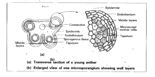
Draw the diagram of a microsporangium and label its wall layers. Write briefly about the wall layers.Solution in Telugu

A typical angiospermics anther is bilobed with each lobe having two theca. The anther is a four sided structure consisting of four microsporangia located at the corners, two in each lobe.
In a transverse section, a typical microsporangium is circular in out line and is surrounded by four wall layers, the a) epidermis b) endothecium, c) walt layers, d) tapetum.
a) Epidermis : The epidermis is one called thick, the cells present between the pollen sacs are the thin walled and their region is called as stomium which is useful for the dehiscence of pollen sacs.
b) Endothecium : Itis present below the epidermis and expands radically with fibrous thickenings, at maturity these cells loose water and contract and help in the dehiscence of pollen sacs.
c)Wall layers : Beneath the Endothecium, there are thin walled cells, arranged in one to five layers, which also help in dehiscence of Anther.
d) Tapetum : The innermost wail layer is Tapetum, the cells are large, with thin cell walls, abundant cytoplasm and have more than one nuclei. Tapetum is a nutritive tissue which nourishes the developing pollen grains.
The centre of the microsporengium consists of sporogeneous tissue, which undergo meiotio divisions to form microspore tetrads. This process is known as Microsporogenesis.
Topper's Solved these Questions
- SEXUAL REPRODUCTION IN FLOWERING PLANTSBook:VIKRAM PUBLICATION ( ANDHRA PUBLICATION)Chapter:SEXUAL REPRODUCTION IN FLOWERING PLANTSExercise:EXERCISES
Explore 6 Videos
- SEXUAL REPRODUCTION IN FLOWERING PLANTSBook:VIKRAM PUBLICATION ( ANDHRA PUBLICATION)Chapter:SEXUAL REPRODUCTION IN FLOWERING PLANTSExercise:Important questions
Explore 13 Videos
- SEXUAL REPRODUCTION IN FLOWERING PLANTSBook:VIKRAM PUBLICATION ( ANDHRA PUBLICATION)Chapter:SEXUAL REPRODUCTION IN FLOWERING PLANTSExercise:Short answer type questions
Explore 10 Videos
- SCIENCE OF PLANTS - BOTANYBook:VIKRAM PUBLICATION ( ANDHRA PUBLICATION)Chapter:SCIENCE OF PLANTS - BOTANYExercise:IMPORTANT QUESTIONS
Explore 8 Videos
- STRUCTURAL ORGANISATION IN ANIMALS Book:VIKRAM PUBLICATION ( ANDHRA PUBLICATION)Chapter:STRUCTURAL ORGANISATION IN ANIMALS Exercise:Important Question
Explore 24 Videos
Similar Questions
Vivipary automatically limits the number of offsprings in a litter How ?
View SolutionName the component cells of the "egg apparatus" in an embryo sac.
View SolutionMention two strategies evolved to prevent self-pollination in flowers.
View SolutionName the books written Parasara and mention the important aspects discussed in those books.
View SolutionWhat is tissue level of organisation among animals? Which metazoans do exhibit this organisation?
View SolutionThe outermost and innnermost wall layers of microsporangium in an anther are respectively
View SolutionDraw the diagram of a microsporangium and lable its wall layers.Write briefly about the wall layers?
View SolutionThe innermost wall layer of microsporangium is called.
View SolutionThe innermost wall layer of microsporangium is the __________
View SolutionDraw and label the enlarged view of microsporangium of an angiosperm . State the function of its innermost wall layer.
View SolutionDraw a anther lobe and label the wall layers.
View SolutionWrite briefly about the different layers of the human heart muscle wall.
View SolutionDraw the diagram of a microsporangium and lebel its wall parts. Write briefly on role of endothecuium.
View SolutionDraw the diagram of a microsporangium and label its wall layers. Write briefly on the role of the endothecium.
View SolutionThe outermost and innermost wall layers of microsporangium in an anther are respectively :
View Solution

