Pulmonary ventilation movements are due to
costal muscles
diaphragm
wall of lungs
costal muscles and diaphragm
Step-by-Step Solution:
1. Understanding Pulmonary Ventilation:
- Pulmonary ventilation refers to the process of moving air in and out of the lungs. It is essential for gas exchange, allowing oxygen to enter the bloodstream and carbon dioxide to be expelled.
2. Identifying the Mechanisms of Ventilation:
- The primary muscles involved in pulmonary ventilation are the diaphragm and the intercostal muscles. The diaphragm is a dome-shaped muscle located at the base of the thoracic cavity, while the intercostal muscles are located between the ribs.
3. Role of the Diaphragm:
- During inhalation (inspiration), the diaphragm contracts and moves downward, increasing the volume of the thoracic cavity. This creates a negative pressure that draws air into the lungs.
4. Role of Intercostal Muscles:
- The intercostal muscles also play a crucial role. When these muscles contract, they elevate the ribs, further expanding the thoracic cavity and allowing more air to flow into the lungs.
5. Pressure Gradients:
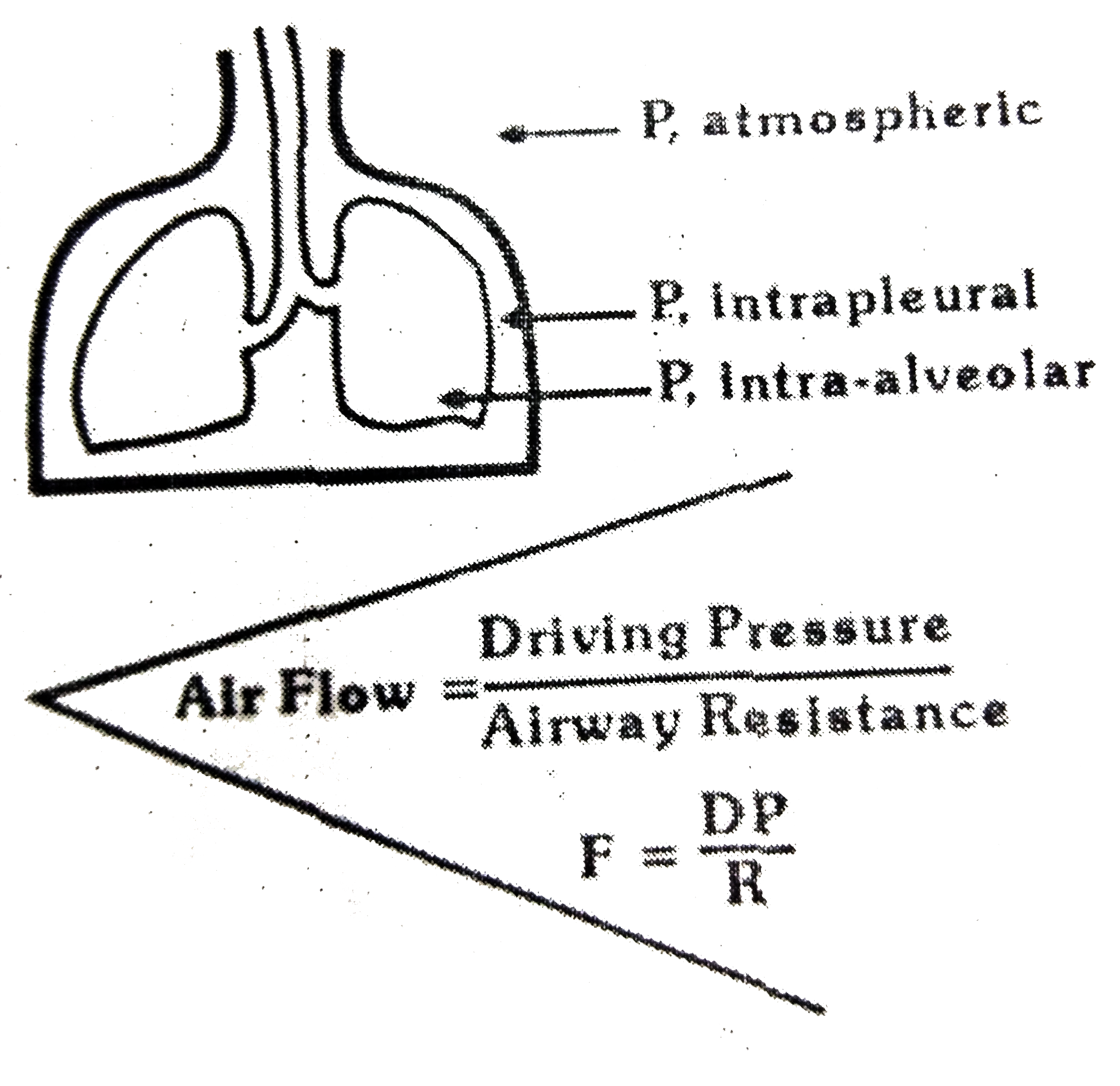
- The movement of air during pulmonary ventilation is driven by pressure gradients. The contraction and relaxation of the diaphragm and intercostal muscles change the pressure within the thoracic cavity, facilitating the flow of air.
6. Conclusion:
- Therefore, pulmonary ventilation movements are due to the combined action of the diaphragm and the intercostal muscles. The correct answer is that pulmonary ventilation is due to both the costal muscles (intercostal muscles) and the diaphragm.
Final Answer:
Pulmonary ventilation movements are due to the costal muscles and diaphragm.
---
- Pulmonary ventilation refers to the process of moving air in and out of the lungs. It is essential for gas exchange, allowing oxygen to enter the bloodstream and carbon dioxide to be expelled.
- The primary muscles involved in pulmonary ventilation are the diaphragm and the intercostal muscles. The diaphragm is a dome-shaped muscle located at the base of the thoracic cavity, while the intercostal muscles are located between the ribs.
- During inhalation (inspiration), the diaphragm contracts and moves downward, increasing the volume of the thoracic cavity. This creates a negative pressure that draws air into the lungs.
- The intercostal muscles also play a crucial role. When these muscles contract, they elevate the ribs, further expanding the thoracic cavity and allowing more air to flow into the lungs.
- The movement of air during pulmonary ventilation is driven by pressure gradients. The contraction and relaxation of the diaphragm and intercostal muscles change the pressure within the thoracic cavity, facilitating the flow of air.
- Therefore, pulmonary ventilation movements are due to the combined action of the diaphragm and the intercostal muscles. The correct answer is that pulmonary ventilation is due to both the costal muscles (intercostal muscles) and the diaphragm.
Pulmonary ventilation movements are due to the costal muscles and diaphragm.
Topper's Solved these Questions
- BREATHING AND EXCHANGE OF GASESBOOK - ICSECHAPTER - BREATHING AND EXCHANGE OF GASESEXERCISE - REVIEW QUESTIONS (FILL IN THE BLANKS)
13 Videos
- BREATHING AND EXCHANGE OF GASESBOOK - ICSECHAPTER - BREATHING AND EXCHANGE OF GASESEXERCISE - REVIEW QUESTIONS (MATCH THE FOLLOWING)
1 Videos
- BODY FLUIDS AND CIRCULATIONBOOK - ICSECHAPTER - BODY FLUIDS AND CIRCULATIONEXERCISE - DIFFRENTIATE BETWEEN
13 Videos
- CELL - THE UNIT OF LIFEBOOK - ICSECHAPTER - CELL - THE UNIT OF LIFEEXERCISE - REVIEW QUESTIONS (LONG ANSWER TYPE QUESTIONS)
14 Videos
Similar Questions
Knowledge Check
The ventilation movements of the lungs in mammals are governed by
Amuscular walls of lungBdiaphramCcostal musclesDboth (b) and (c)

The alveolar ventilation per minute refers to the amount of fresh air which reaches the alveoli of the lungs per minute. Alvelor ventilation per minute equals the
View SolutionThe ventilation movements of the lungs in mammals are governed by
View SolutionPhototropism is shoots is attributed to or photoropic movements are due to
View SolutionStudy the diagram given below and answer the questions that follow:

Which stage of pulmonary ventilation is shown in this diagram ? Give reason in support of your answer.
View SolutionThe alveolar ventilation is the
View SolutionVentilation movements in Cockroach are performed
View SolutionAssertion : Maximum movement is possible at the amphiarthrosis joint.
Reason : Such joint are also called synovial joints and have almost frictionless movement due to synovial fluid.
View Solution
