Draw a vertical section of a maize grain and label (i) pericarp, (ii)scutellum, (iii)coleoptile, and (iv) radicle
To draw a vertical section of a maize grain and label the specified parts, follow these steps:
1. Draw the Outline of the Maize Grain:
- Begin by sketching the general shape of a maize grain. It resembles a slightly elongated oval or a corn kernel.
2. Draw the Pericarp:
- The pericarp is the outer layer of the grain. Draw a thin line around the outer edge of the grain to represent the pericarp. This layer protects the seed.
3. Draw the Scutellum:
- Inside the grain, draw a flat, shield-like structure that sits against the endosperm. This is the scutellum, which is responsible for nutrient absorption during germination.
4. Draw the Coleoptile:
- Above the scutellum, draw a small, elongated structure that looks like a protective sheath. This is the coleoptile, which will protect the emerging shoot as it grows upward.
5. Draw the Radicle:
- Below the scutellum, draw a small, pointed structure that extends downward. This is the radicle, which will develop into the root of the plant.
6. Label the Parts:
- Clearly label each part of the maize grain:
- "Pericarp" for the outer layer,
- "Scutellum" for the shield-like structure,
- "Coleoptile" for the protective sheath above the scutellum,
- "Radicle" for the root structure below the scutellum.
Final Diagram:
- Your final drawing should show the vertical section of the maize grain with all parts labeled clearly.
- Begin by sketching the general shape of a maize grain. It resembles a slightly elongated oval or a corn kernel.
- The pericarp is the outer layer of the grain. Draw a thin line around the outer edge of the grain to represent the pericarp. This layer protects the seed.
- Inside the grain, draw a flat, shield-like structure that sits against the endosperm. This is the scutellum, which is responsible for nutrient absorption during germination.
- Above the scutellum, draw a small, elongated structure that looks like a protective sheath. This is the coleoptile, which will protect the emerging shoot as it grows upward.
- Below the scutellum, draw a small, pointed structure that extends downward. This is the radicle, which will develop into the root of the plant.
- Clearly label each part of the maize grain:
- "Pericarp" for the outer layer,
- "Scutellum" for the shield-like structure,
- "Coleoptile" for the protective sheath above the scutellum,
- "Radicle" for the root structure below the scutellum.
- Your final drawing should show the vertical section of the maize grain with all parts labeled clearly.
Topper's Solved these Questions
- REPRODUCTIONBook:TRUEMEN BIOLOGY ENGLISHChapter:REPRODUCTIONExercise:SECTION -C
Explore 22 Videos
- REPRODUCTIONBook:TRUEMEN BIOLOGY ENGLISHChapter:REPRODUCTIONExercise:SECTION-D
Explore 27 Videos
- REPRODUCTIONBook:TRUEMEN BIOLOGY ENGLISHChapter:REPRODUCTIONExercise:SECTION-D
Explore 27 Videos
- Protista Book:TRUEMEN BIOLOGY ENGLISHChapter:Protista Exercise:ASSERTION AND REASON
Explore 6 Videos
- Reproduction In OrganismsBook:TRUEMEN BIOLOGY ENGLISHChapter:Reproduction In OrganismsExercise:MULTIPLE CHOICE QUESTION
Explore 131 Videos
Similar Questions
Draw a diagram of sectional view of human ovary and label (i) Primary follicle, (ii) Tertiary follicle, (iii) Graafian follicle and (iv) Corpus luteum.
View SolutionDraw a diagram of the vertical section of human eye and label the following parts : (i) cornea, (ii) iris, (iii) aqueous humour, (iv) retina, (v) ciliary body, (vi) fovea centralis, (vii) choroid, (viii) pupil, and (ix) lens.
View Solution(a) Draw a diagram of Pistil showing pollen tube growth in angiosperm and label (i) Stigma, (ii) male gametes , (iii) micropyle and (iv) Ovule.
(b) Write the function of micropyle.
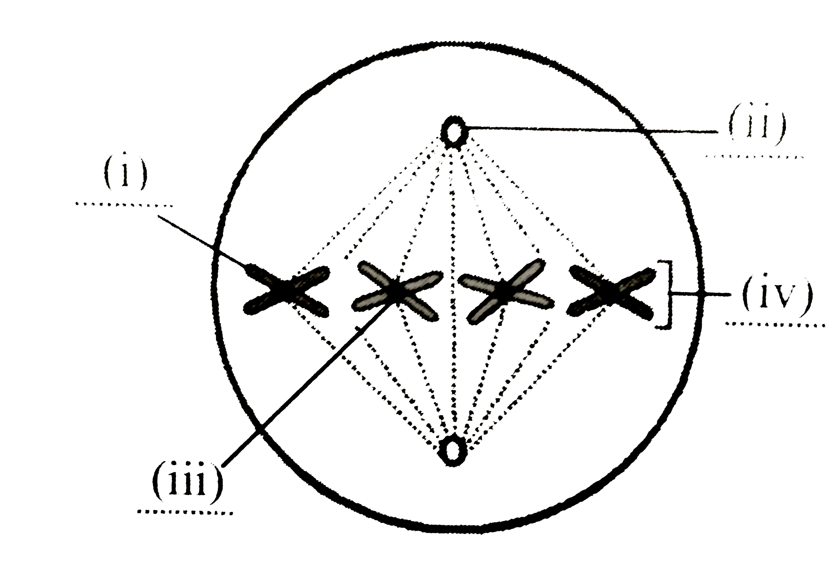
View SolutionLabel the parts (i), (ii), (iii) and (iv)

View SolutionDraw the structure of the human ear and label the following parts: (i) tympanum, (ii) cochlea, (iii) pinna, (iv) malleus, (v) semi-circular canals, (vi) eustachian tube.
View SolutionDraw a vertical section of the human eye and label any six of those parts only through which the light rays pass and fall on the retina.
View SolutionDraw a diagram of the vertical section of human eye and label any six parts surrounding the vitreous chamber.
View Solution(a) Draw a diagrammatic sectional view of a seminiferous tubule (enlarged) of human male and label (i) Spermatogenium, (ii) Sertoli cell, (iii) Primary spermatocyte. Write the function of each labelled part.
(b) Differentiate between spermatogenesis and spermiogenesis.
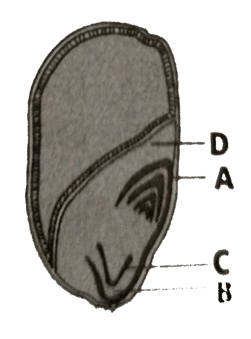
View SolutionGiven figure represents longitudinal section of a monocotyledonous embryo.
Identify the parts labelled as A,B, C and D from the list (i-vii) and select the correct option

(i) Scutellum
(ii) Coleoptile
(iii) Shoot apex
(iv) Epiblast
(v) Radicle
(vi) Root Cap
(vii) Colerhiza
View SolutionLabel the structure indicated by (i), (ii), (iii) and (iv)-

View Solution

