The given diagram shows L.S. of testis showing various parts. Identify the parts labelled (A to G) from the list given below.
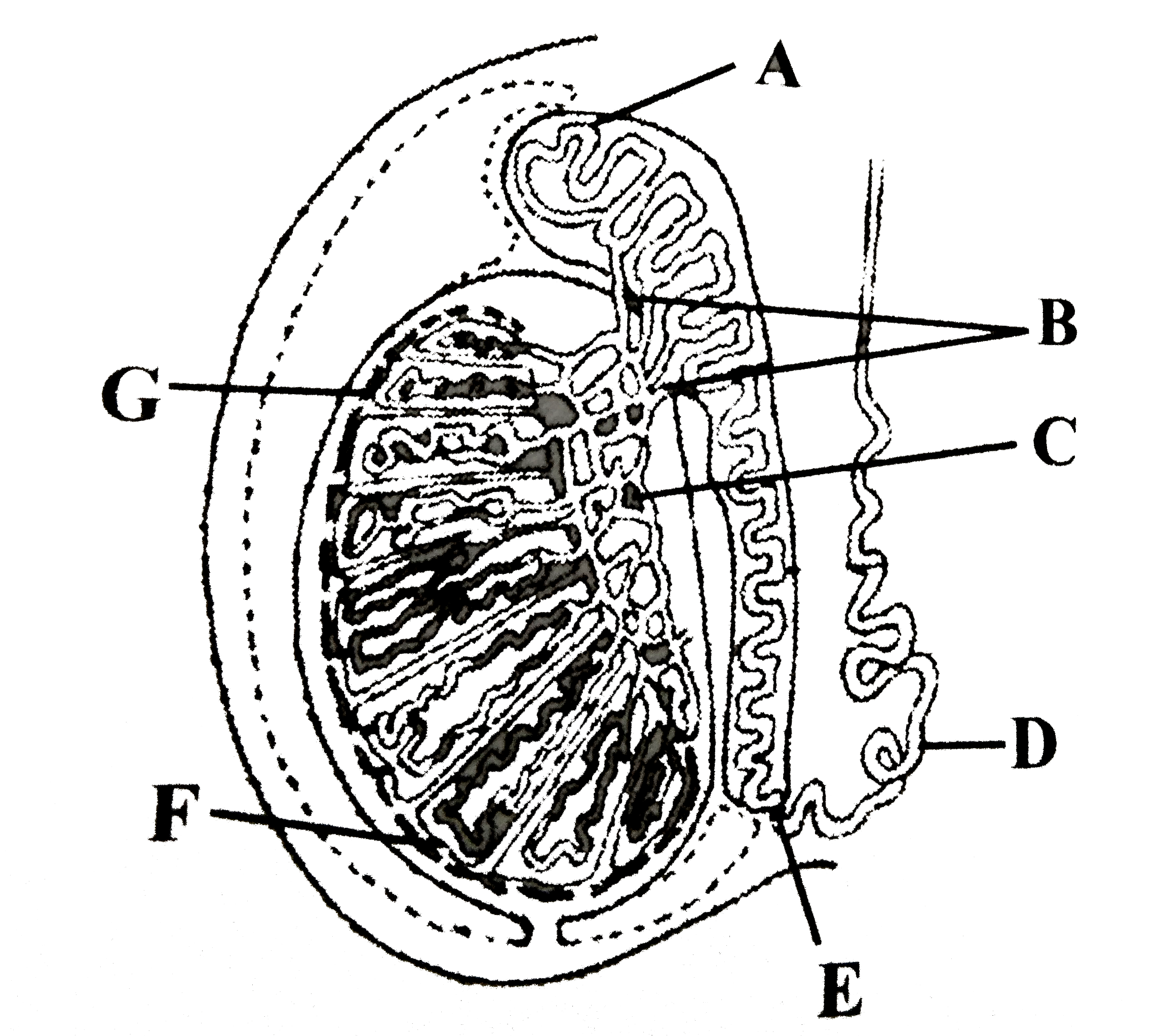
(i)Caput epididymis(ii)Cauda epididymis(iii)Vas deferens(iv)Vasa efferentia(v)Corpus epididymis(vi)Seminiferous tubulus(vii)Tunica vaginalis(Viii)Tunica albuginea(ix)Tunica vasculosa(x)Rate testis
A−(ii),B−(iii),C−(iv),D−(x),E−(vii),F−(i),G−(ix)
A−(v),B−(iv),C−(iii),D−(vi),E−(i),F−(x),G−(vii)
A−(i),B−(iv),C−(x),D−(iii),E−(ii),F−(vi),G−(viii)
A−(i),B−(vi),C−(iv),D−(iii),E−(v),F−(x),G−(ix)
The correct Answer is:C
Step by step video, text & image solution for The given diagram shows L.S. of testis showing various parts. Identify the parts labelled (A to G) from the list given below. {:((i),"Caput epididymis",(ii),"Cauda epididymis"),((iii),"Vas deferens",(iv),"Vasa efferentia"),((v),"Corpus epididymis",(vi),"Seminiferous tubulus"),((vii),"Tunica vaginalis",(Viii),"Tunica albuginea"),((ix),"Tunica vasculosa",(x),"Rate testis"):} by Biology experts to help you in doubts & scoring excellent marks in Class 12 exams.
Updated on:21/07/2023Topper's Solved these Questions
- HUMAN REPRODUCTIONBook:NCERT FINGERTPS ENGLISHChapter:HUMAN REPRODUCTIONExercise:Human Reproduction
Explore 185 Videos
- HUMAN HEALTH AND DISEASEBook:NCERT FINGERTPS ENGLISHChapter:HUMAN HEALTH AND DISEASEExercise:Human Health And Disease
Explore 177 Videos
- MICROBES IN HUMAN WELFAREBook:NCERT FINGERTPS ENGLISHChapter:MICROBES IN HUMAN WELFAREExercise:Microbes In Human Welfare
Explore 138 Videos
Similar Questions
Given figure represents longitudinal section of a monocotyledonous embryo.
Identify the parts labelled as A,B, C and D from the list (i-vii) and select the correct option
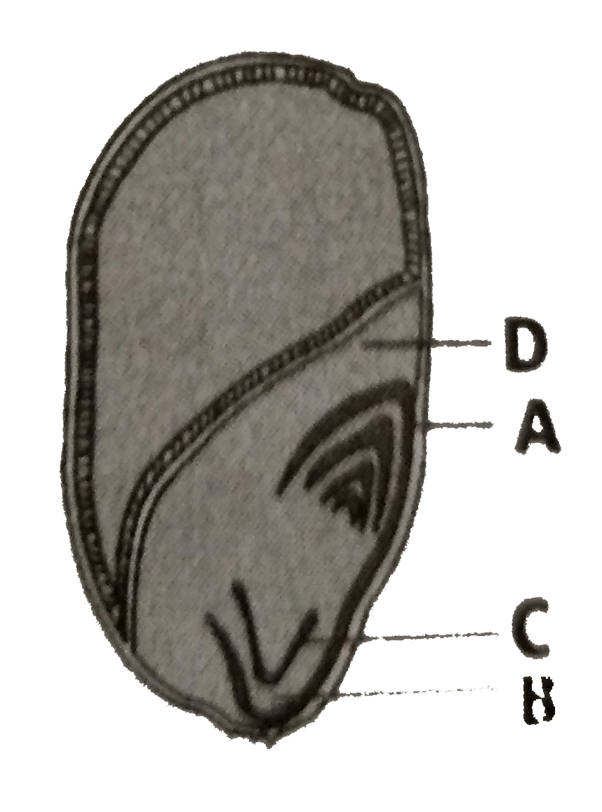
(i) Scutellum
(ii) Coleoptile
(iii) Shoot apex
(iv) Epiblast
(v) Radicle
(vi) Root Cap
(vii) Colerhiza
View SolutionMatch column I with column II and select the correct option from the codes given below
Column IColumn IIA. Lungs(i). Lactic acidB. Liver(ii). Hypertonic urineC. Micturition(iii). Counter-current systemD. Sweat(iv). CO2E. Vasa recta(v). Urinary bladderF. Sebum(vi). GlucoseG. ADH(vii). BilirubinH. Tubular reabsorption(viii). Sterols
View Solution
Knowledge Check
The given figure shows a section of brain. Identify the parts labelled as A,B,C and D and match them with the names (i-vii) given below.
(i)Arachnoid membrane , (ii)Subdural space , (iii)Duramater , (iv)Bone , (v)White matter , (vi)Grey matter , (vii)Piamater
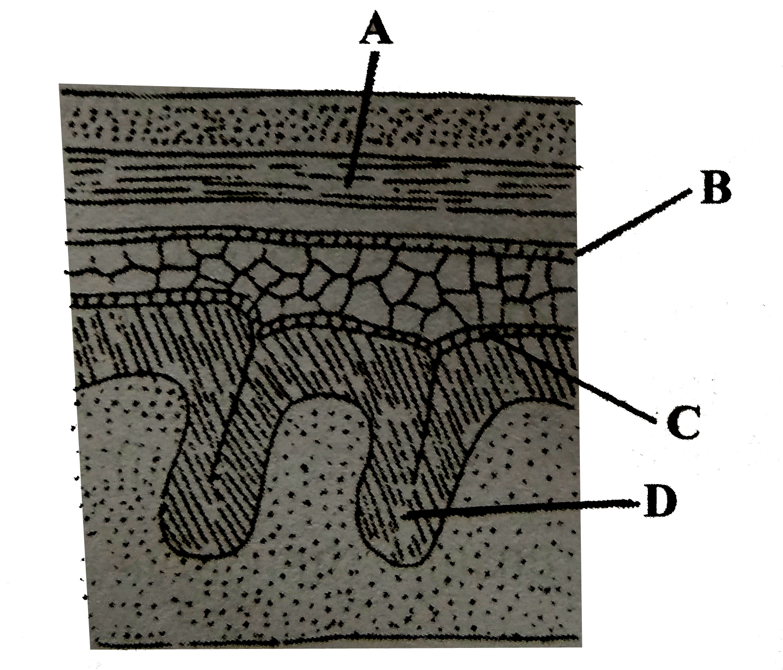 AABCD(iii)(ii)(vi)(v)BABCD(i)(ii)(iii)(vi)CABCD(iii)(i)(vii)(vi)DABCD(iv)(vii)(i)(ii)
The given figure is of circulatory system. Identify the labelled parts (A-D) from the list (i-vii).
(i) Pulmonary circulation
(ii) Systemic circulation
(iii) Superior vena cava
(iv) Inferior vena cava
(v) Aorta
(vi) Veins and venules
(vii) Arterioles and capillaries
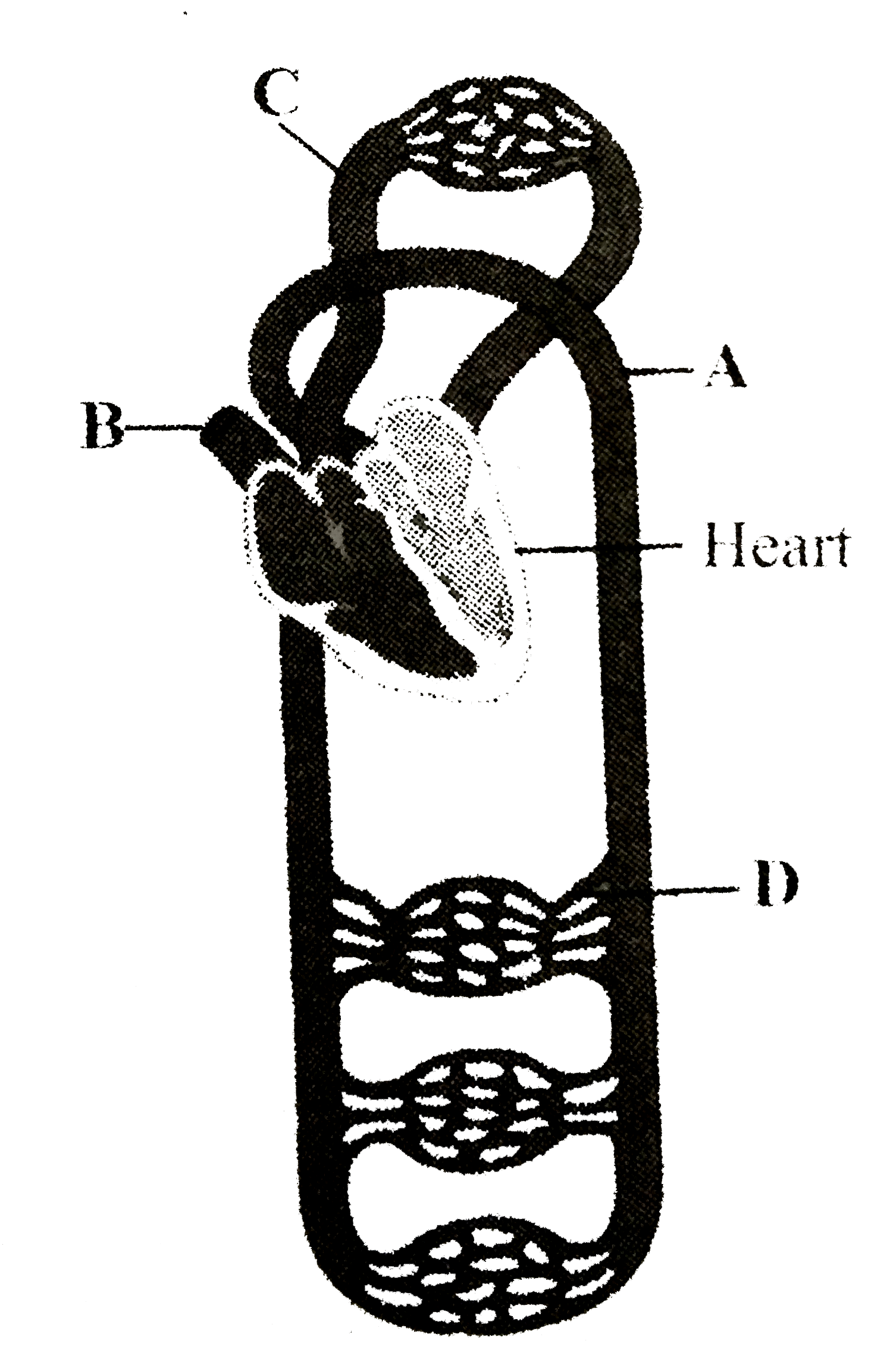 AA B C D(v) (iii) (i) (vii)BA B C D(vii) (iv) (i) (vi)CA B C D(v) (iii) (ii) (vii)DA B C D(vii) (v) (i) (vi)
Identify the parts labelled as A,B,C and D in the given figure and match the correct names from the list (i-viii) given below.
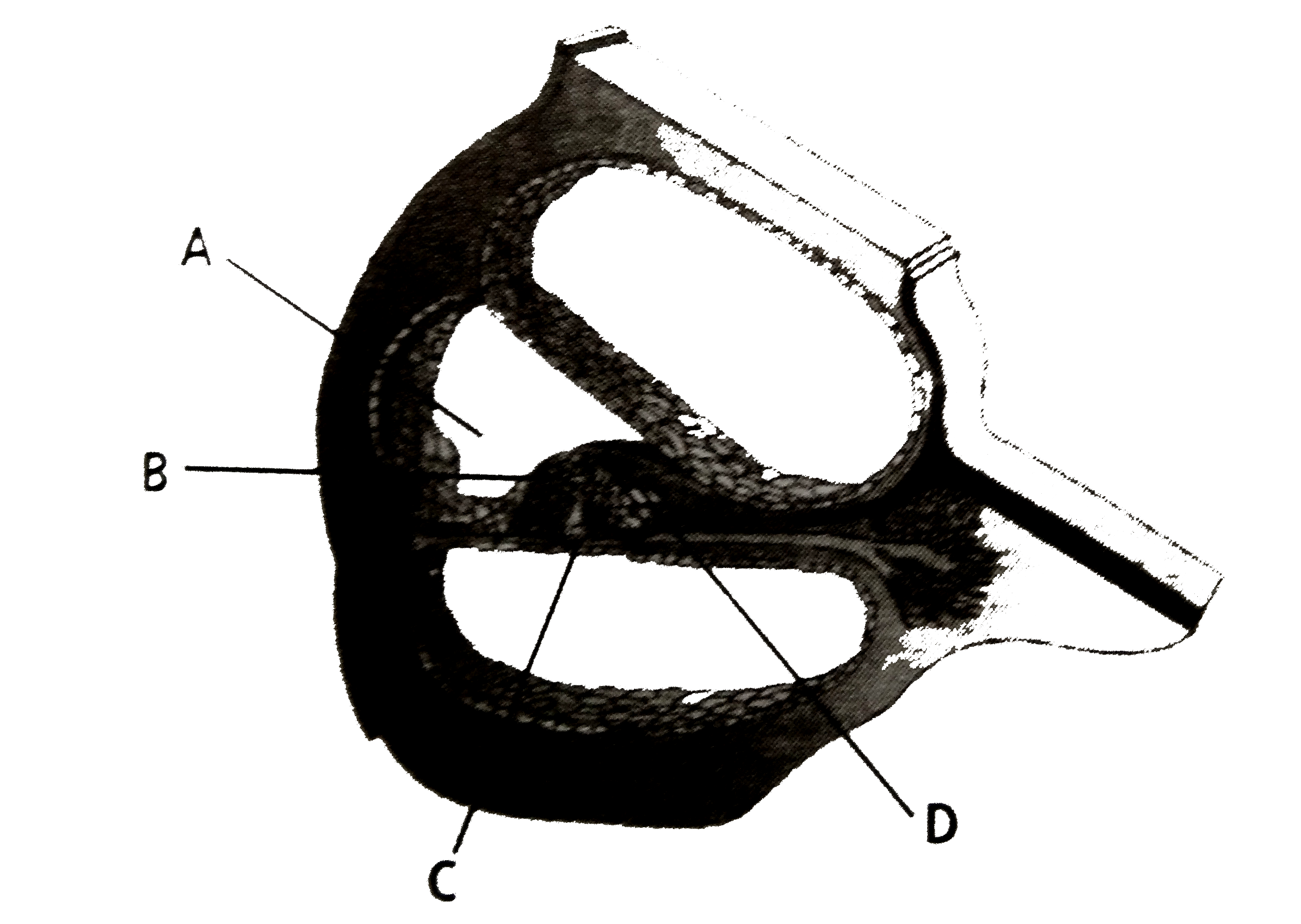
(i)Reissner's membrane , (ii)Basilar membrane
(iii)Tectorial membrane , (iv)Organ of Corti
(v)Hair cellls , (vi)Otolith organ
(vii)Scala media , (viii)Scala vestibuliAABCD(ii)(v)(iii)(i)BABCD(vii)(iv)(ii)(iii)CABCD(vii)(iv)(i)(ii)DABCD(viii)(vi)(i)(iii)
Given below is the outline of the cross-section of the male reproductive system :

(i) Copy the outline on to your answer sheet with pencil and label the following parts testis, epididymis, seminal vesicles, vas deferens
(ii) Name the hormone produced by the testis.
(iii) Why are sperms produced in large numbers?
(iv) State the function of the seminal vesicles.
View SolutionIdentify the components labelled A, B, C and D in the diagram below from the list (i) to (viii) given along with
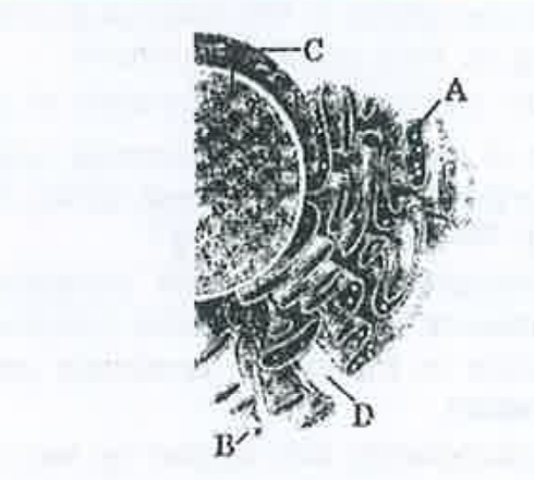
Components :
(i) Cristae of mitochondria
(ii) Inner membrane of mitochondria
(iii) Cytoplasm
(iv) Smooth endoplasmic reticulum
(v) Rough endoplasmic reticulum
(vi) Mitochondrial matrix
(vii) Cell vacuole
(viii) Nucleus
The correct components are
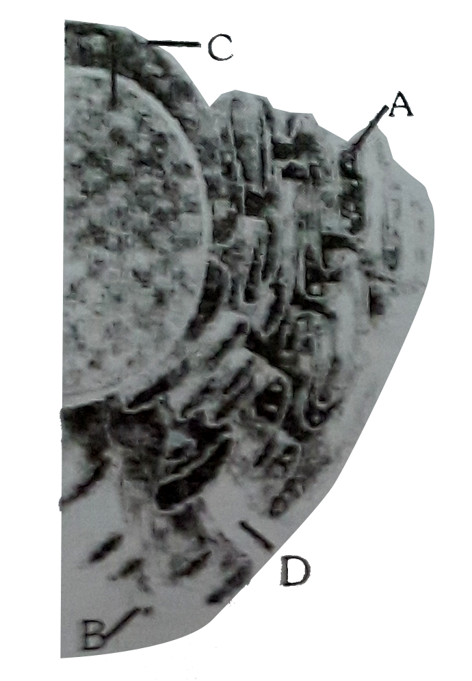
View SolutionIdentify the components labelled A, B, C and D in the diagram below from the list (i) to (vii) given along with
Components :
(i) Cristae of mitochondria
(ii) Inner membrane of mitochondria
(iii) Cytoplasm
(iv) Smooth endoplasmic reticulum
(v) Rough endoplasmic
(vi) Mitochondrial matrix
(vii) Cell vacuole
(viii) Nucleus
The correct components are
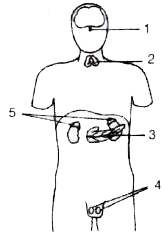
View SolutionThe given diagram is an outline of human body showing the posttion of certain organs. In this diagram
(I) Name the parts labelled 1-5.
(ii) Name the hormone released from 1 that influences the working of part 2.
(iil) How do part labelled as 3 differs from parts labelled as 1 and 2 in general? Name the endocrine cells present in part 3.
(iv) What will happen if the part labelled as 4 is removed ?
(11) Name the hormones released by part 5.
(VI) Why the part labelled as 1 called master gland? Which part of the forebrain controls the gland labelled 1 ?
(vii) Name the gland that secretes 'emergency hormone.
View SolutionThe given diagram shows axon terminal.Select the option that correctly matches the steps in transmission of impulses (list i-vii) with the labellings (A-C) in diagram.
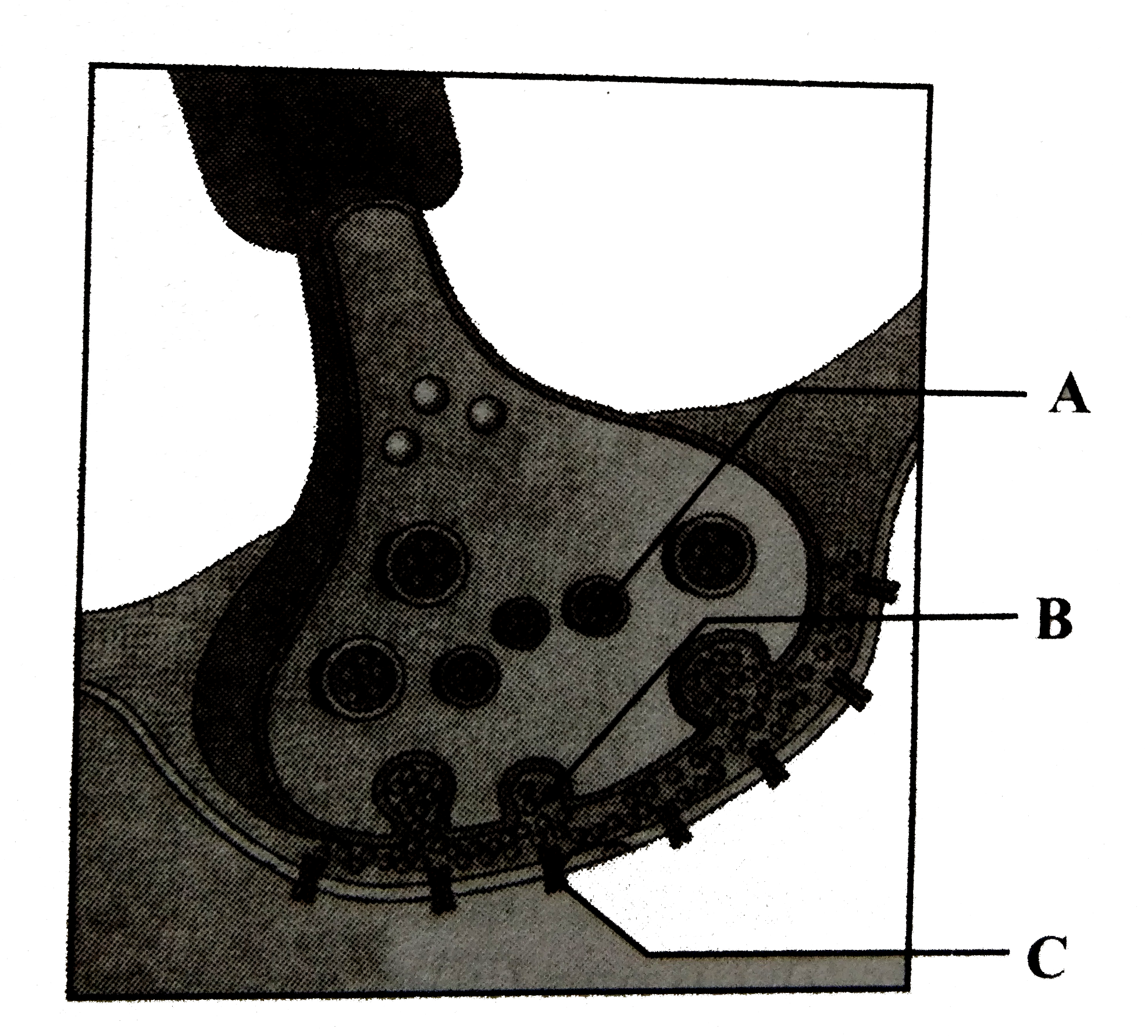
(i)Chemicals called neurotransmitters are released in the synaptic cleft through ion channels.
(ii)When an impulse arrives at the axon terminal, it stimulates the movement of synaptic vesicles.
(iii)Neurotransmitters are endocytosed into the neurons.
(iv)The ions channels close with the binding of neurotransmitters to their specific receptors vesicles.
(v)Synaptic vesicles move towards the membrane where they fuse with the plasma membrane.
(vi)Neurotransmitters are released in the synaptic cleft.
(vii)The released transmitters bind to their specific receptors on post-synaptic membrane
View Solution

