Explain the structure of blood vessels. Solution in Tamil
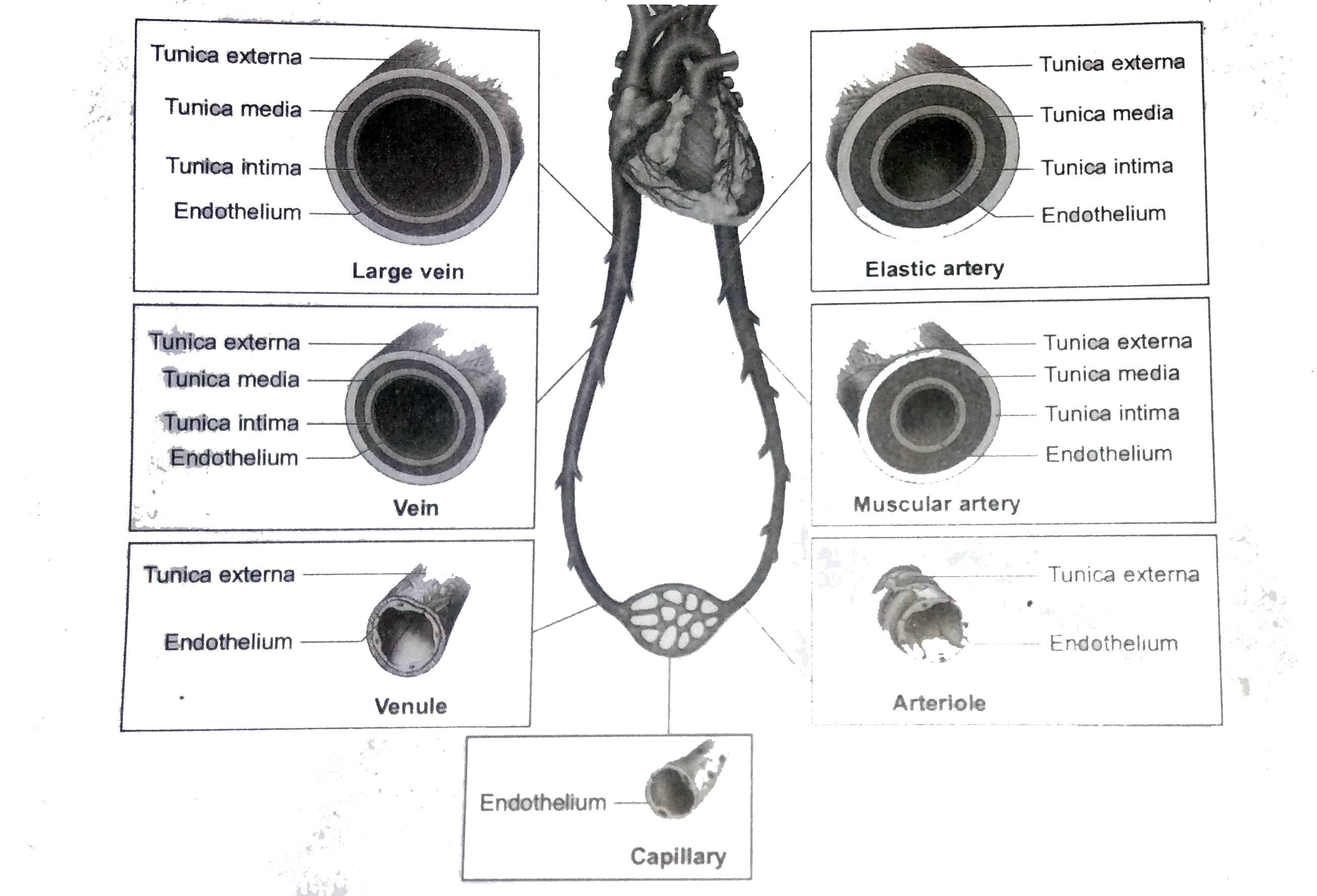
The vessels carrying the blood are of three types, they are the arteries, veins and capillaries. These vessels are hollow structures and have complex walls surrounding the lumen. The blood vessels in humans are composed of three layers, tunica intima, tunica media and tunica externa. The inner layer, tunica intima or tunica interna supports the vascular endothelium, The middle layer, tunica media is composed of smooth muscles and an extra cellular matrix wcn contains a protein, elastin. The contraction and relaxation of the smooth muscles results vsoconstriction and vasodilation. The outer layer, tunica externa or tunica adventitia is composed of collagen fibres.
Arteries: The blood vessels that carry blood away from the heart are called arteries. T he aiCs usualy lhe deep inside the body. The walls of the arteries are thick, non-collapsible to withstand high pressure. Valves are absent and have a narrow lumen. All arteries carry Oxygenated blood, except the pulmonary artery. The largest artery, the aorta (2.5 cm in damere and 2 mm thick) branch into smaller arteries and culminates into the tissues as TeCed arci n the tissues the arteries branches into arterioles. As blood enters an arteriole it may have a pressure of 85 mm Hg (11.3 KPa) but as it leaves and flows into the capilary, the pressure drops to 35 mm Hg (4.7 KPa). (Note 1 mm Hg = 0.13 KPa. SI unit of mm Hg is KiloPascal (KPa)). Arterioles are small, narrow, and thin walled which are connected to the capillaries. A Small sphincter lies at the junction between the arterioles and capillaries to regulate the blood supply. Arteries do not always branch into arterioles, they can also form anastomoses.

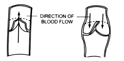
Capillaries: Capillary beds are made up of fine networks of capillaries. The capillaries are thin walled and consist of single layer of squamous epithelium. Tunica media and elastin fibres are absent. The capillary beds are the site for exchange of materials between blood and tissues. The walls of the capillaries are guarded by semilunar valves. The blood volume in the capillaries is high but the flow of blood is slow. Mixed blood (oxygenated and deoxygenated) is present in the capillaries. The capillary bed may be fiooded with blood or may be completely bypassed depending on the body conditions in a particular organ. Veins: Veins have thinner walls and a larger lumen and hence can be easily stretched. They carry deoxygenated blood except, the pulmonary vein. The blood pressure is low and the lumen has a wide wall which is collapsible. Tunica media is thinner in veins than in arteries. Unidirectional flow of blood in veins is due to the presence of semilunar valves that prevents backflow of blood. Blood samples are usually taken from the veins rather than artery because of low pressure in the veins.
Topper's Solved these Questions
- BODY FLUIDS AND CIRCULATIONBOOK - FULL MARKSCHAPTER - BODY FLUIDS AND CIRCULATIONEXERCISE - ADDITIONAL QUESTIONS SOLVED ( Fill in the Blanks)
76 Videos
- BIOMOLECULESBOOK - FULL MARKSCHAPTER - BIOMOLECULESEXERCISE - ADDITIONAL QUESTIONS SOLVED (Higher Order Thinking Skills)
16 Videos
- CELL CYCLEBOOK - FULL MARKSCHAPTER - CELL CYCLEEXERCISE - ADDITIONAL QUESTIONS SOLVED (Higher Order Thinking Skills)
12 Videos
Similar Questions
The tissue fluid that surrounds the cell is ........
View SolutionIn which form does the glucose is stored in animal cells? Specify the cells?
View SolutionGiven that the average duplication time of E. coli is 20 minutes. How much time will two E. coli cells takes to become 32 cells?
View Solutionबोमेन सम्पुट में रक्त ले जाने वाली वाहिनी है
View Solutionरक्त परिसंचरण में रक्त वाहिनियों की भूमिका बताइए।
View SolutionPlant structure comparable to human blood vessels
View SolutionDraw the the structure of Blood Vessel - Large Vein.
View SolutionCirculatory system-Blood and Blood vessels
View SolutionCommisural blood vessel connects
(A) Dorsal blood vessel to ventral blood vessel
(B) Sub neural blood vessel to Dorsal blood vessel
(C) Sub neural blood vessel to supro oesophageal blood vessel
(D) Latero oesophageal blood vessel to supro oesophageal blood vessel
View SolutionBlood vessels in the blood vessels are not caused by the presence of
View SolutionThe diagram given below represents a certain category of blood vessels showing the role of a special structure in their walls :
 Describe the role of these structures.
View SolutionThe diagram given below represents a certain category of blood vessels showing the role of a special structure in their walls : Are these structures present in any other kind of blood vessel ? If so, name it.
View SolutionExplain the location and functions of various valves present in the heart and blood vessels.
View SolutionHeart, Blood vessels, Blood, Kidney
View SolutionThe blood vessel that gathers blood in earthworm
View Solution
