To study pollen germination, some pollen from flowers such as pea, chickpea, Crotolaria, balsam and Vinca are collected and dusted on a glass slide containing a drop of ..a.. Solution about..b..per cent . After about ..c.. Minutes, pollen tubes coming out of the pollen grains.
a - saline, b - 5, C - 5 to 10
a - sugar, 6-5, C - 10 to 20
a - saline, b - 10, C - 15 to 30
a - Sugar, b - 10, C - 15 to 30
The correct Answer is:4
Step by step video & text solution for To study pollen germination, some pollen from flowers such as pea, chickpea, Crotolaria, balsam and Vinca are collected and dusted on a glass slide containing a drop of ..a.. Solution about..b..per cent . After about ..c.. Minutes, pollen tubes coming out of the pollen grains. by Biology experts to help you in doubts & scoring excellent marks in Class 11 exams.
Updated on:21/07/2023Similar Questions
Draw a longitudinal section of the pistil from a flowering plant, where pollination has occurred. Label the following: (a) Stigma showing germinating pollen grains (b) Style (c) Pollen tube reaching the micropyle of the ovule (d) Embryo sac (e) Components of the egg apparatus
View Solution(a) Draw a diagram showing germination of pollen on stigma of a flower and mark on it the following organs/parts:
(i) Pollen Grain
(ii) Pollen tube
(iii) Stigma
(iv) Female germ cell
(b) State the significance of pollen tube.
(c ) Name the parts of flower that develop after fertilisation into
(i) Seed (ii) Fruit
View Solution
Knowledge Check
Even after killing the generative cell with a laser beam, the pollen grain of a flowering plant germinates and produces normal pollen tube because
ALaser beam stimulates pollen germination and pollen tube growthBLaser beam does not damage the region from which pollen tube emergesCThe contents of the killed generative cell permit germination and pollen growthDThe vegetative cell has not been damagedIf many pollen tubes come out of a pollen grain during its germination, the condition is regarded as
APolygamousBPolyandrousCPolysiphonousDPolytrichousEven after killing the generative cell with a laser beam, the pollen grain of a flowering plant germinates and produces normal pollen tube because
ALaser beam stimulates pollen germination and pollen tube growthBThe laser beam does not damage the region from which pollen tube emergiesCThe contents of killed generative cell permit germination and pollen tube growthDThe vegetative cell has not been damaged
Which of the following is an outbreeding device ?
a. If pollen release and stigma receptivity are not synchronised.
b. If the anther and stigma are placed at different positions so that pollen cannot come in contact with the stigma of the same flower.
c. Self-incompatibilty which prevents self-pollen (from the same plant) from fertilising the ovules by inhibiting pollen germination or pollen tube growth in the pistil.
d. Production of the unisexual flower
View SolutionTransfer of pollen grains to the stigma of the pistil is termed as .......a........ .
II. Transfer of pollen grains from the anther to the stigma of the same flower is termed as ..........b.......
III . Transfer of pollen grains from the anther to the stigma of another flower of the same plant is termed as ........c.........
Transfer of pollen grains from the anther to the stigma of a different plant is termed as .........d......
Fill in the blanks a, b, ,c and d with given terms .
View SolutionConsider the following statements with respect to the flowering plants and choose the correct option (a) Pollen grains represent the male gametes (b) Functional megaspore develops innto embryo sac that represents the female gamete (c) Transfer of pollen grains from anther to stigma of different plant is known as xenogamy (d) Transfer of pollen grains from anther to stigma of another flower of the same plant is known as geitonogamy
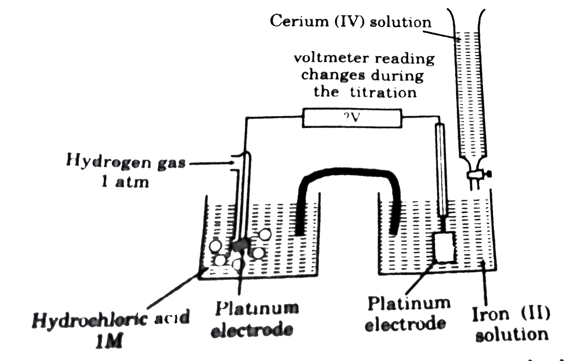
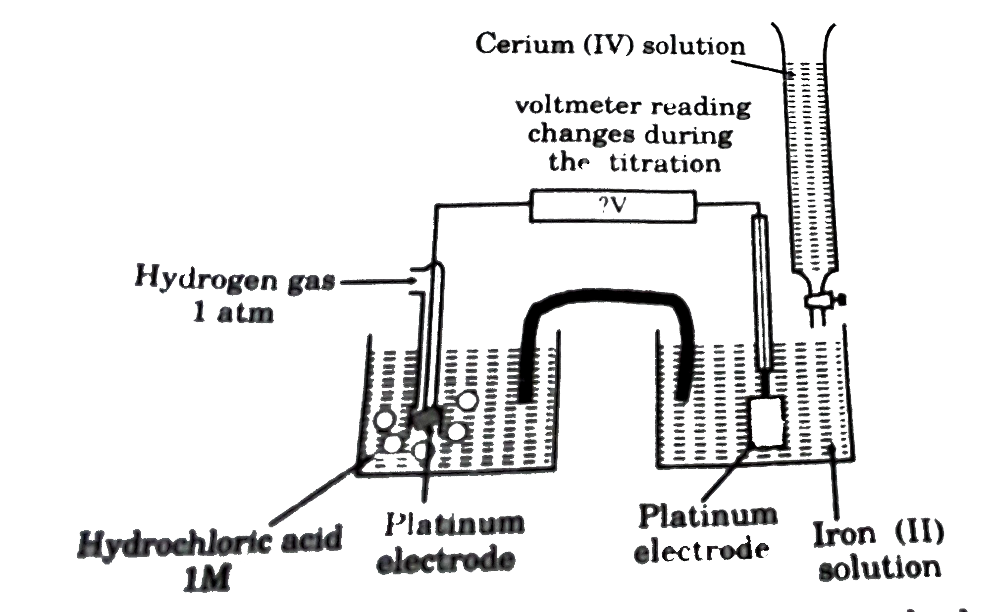
View SolutionTitration are one of the methods we can use to discover the precise concentration of solutions. A typical titration involves adding a solution from a burette to another solution in a flask. The end point of the titration is found by watching a colour change taking place.However, a problem arises when a suitable indicator cannot be found, or when the colour change involved are unclear. In these cases, redox potential may sometimes come to the rescue.

A perticularly well known example (Fig.) is a method of discovering then concentration of iron (II) ions in a solution by titrating them with a solution of cerium (IV). The redox potential that are of interest here areE∘Fe3+∣Fe2+=0.77VandE∘Ce4+∣Ce3+=1.61V. These tell us that cerium (IV) ions are the oxidising agents. They should react according to the equation
Fe2+(aq)+Ce4+(aq)→Fe3+(aq)+Ce3+(aq).
Now imagine that we know the concentration of the cerium (IV) ions solution in the burette. We want to measure the concentration of the cerium (IV) solution from the burette, some of the iron (II) ions are will be oxidised. As a consequence the beaker would now contain large number of unreacted iron (II) ions , but also some iron (III) ions as well. All of the cerium (III). The solution in the beaker now represents an iron (III)/iron(II) half cell although not at standard conditions.Thus , the emf of the cell will be near, but not equal to E∘Fe3+∣Fe2+.

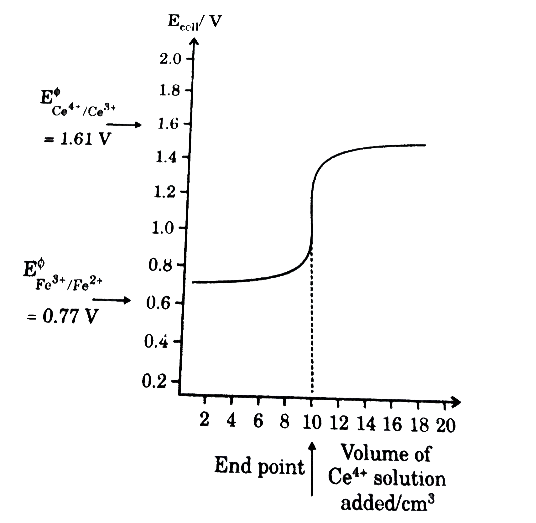
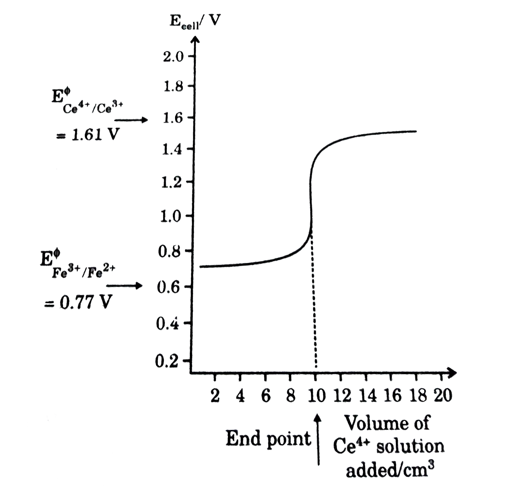
If we continue to add cerium (IV) solution , the number of iron (II) ions is gradually reduced and eventually only a very few are left (Table).At this stage the next few drops of cerium (IV) solution convert all the remaining irom (II) ions into iron (III), and some of the cerium (IV) ions are left unreacted. Once this happens we no longer have an iron (III)/iron(II) half-cell. Instead we have a solution in which there is a large number of cerium (III) ions and a smaller number of cerium (IV) ions. The solution in the beaker now behaves as a cerium (IV)/cerium(III)half-cell (although not a standard one.)
Just before all the iron (II) ions are converted into iron (III) we have a cell with an EMF of around +0.77V. After all the iron (II) ions are oxidised we have a cell with an emf of about +1.61V. This rapid rise in emf occurs with the addition of just one drop of cerium (IV) solution. You should be able to understand why a graph of cell emf against volume of cerium (IV) solution of the iron (II) solution calculated in the usual way.
Imagine you were given a solution of potassium dichromate (VI) in a beaker and a solution of iron (II) sulphate in a burette. You do not know the concentration of dichromate (VI) ions , but the concentration of the iron (II) solution is known. Your task it to carry out a redox titration using the two solutions in order to determine the concentration of dichromate (VI) ions. Sketch a graph showing how the emf changes in the course of the above titration.
ECr2O2−7/Cr3+=1.33V,EFe3+/Fe2+=0.77V
View SolutionTitration are one of the methods we can use to discover the precise concentration of solutions. A typical titration involves adding a solution from a burette to another solution in a flask. The end point of the titration is found by watching a colour change taking place.However, a problem arises when a suitable indicator cannot be found, or when the colour change involved are unclear. In these cases, redox potential may sometimes come to the rescue.

A perticularly well known example (Fig.) is a method of discovering then concentration of iron (II) ions in a solution by titrating them with a solution of cerium (IV). The redox potential that are of interest here areE∘Fe3+∣Fe2+=0.77VandE∘Ce4+∣Ce3+=1.61V. These tell us that cerium (IV) ions are the oxidising agents. They should react according to the equation
Fe2+(aq)+Ce4+(aq)→Fe3+(aq)+Ce3+(aq).
Now imagine that we know the concentration of the cerium (IV) ions solution in the burette. We want to measure the concentration of the cerium (IV) solution from the burette, some of the iron (II) ions are will be oxidised. As a consequence the beaker would now contain large number of unreacted iron (II) ions , but also some iron (III) ions as well. All of the cerium (III). The solution in the beaker now represents an iron (III)/iron(II) half cell although not at standard conditions.Thus , the emf of the cell will be near, but not equal to E∘Fe3+∣Fe2+.

If we continue to add cerium (IV) solution , the number of iron (II) ions is gradually reduced and eventually only a very few are left (Table).At this stage the next few drops of cerium (IV) solution convert all the remaining irom (II) ions into iron (III), and some of the cerium (IV) ions are left unreacted. Once this happens we no longer have an iron (III)/iron(II) half-cell. Instead we have a solution in which there is a large number of cerium (III) ions and a smaller number of cerium (IV) ions. The solution in the beaker now behaves as a cerium (IV)/cerium(III)half-cell (although not a standard one.)
Just before all the iron (II) ions are converted into iron (III) we have a cell with an EMF of around +0.77V. After all the iron (II) ions are oxidised we have a cell with an emf of about +1.61V. This rapid rise in emf occurs with the addition of just one drop of cerium (IV) solution. You should be able to understand why a graph of cell emf against volume of cerium (IV) solution of the iron (II) solution calculated in the usual way.
When an ion is converted into a complex ion, the redox potential change. you can see this in the combination of standard emf of the iron (III)/iron(II) system (+0.36V). The cyanide ion is said to stabilize the oxidation state of the iron. If you were to make up a cell
Pt(s)∣∣ ∣∣Fe(CN)3−61mol dm−1(aq),Fe(CN)4−6∣∣ ∣∣∣∣ ∣∣Fe3+(aq)1mol dm−3,Fe2+(aq)∣∣ ∣∣Pt(s)
what would be the emf and what would be the cell reaction?
View Solution
