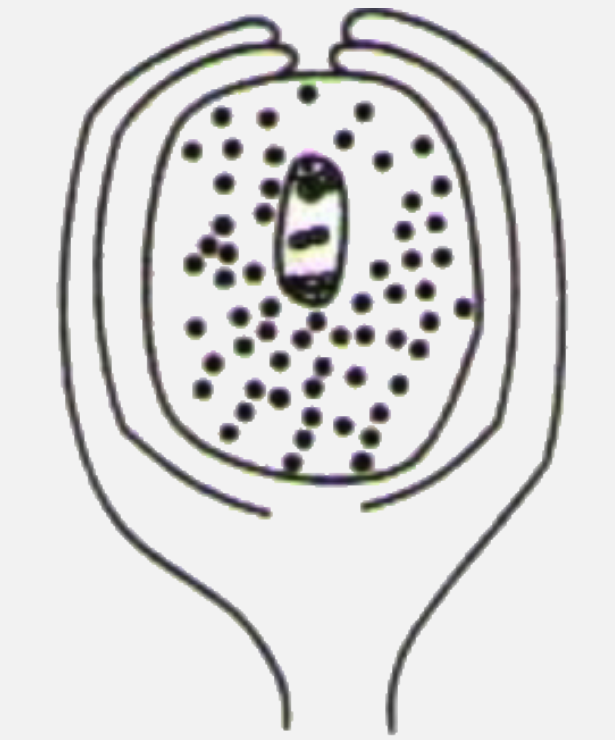
Diagrammatically represent sectional view of trophoblastic organisation in animals.
Step-by-Step Text Solution:
1. Understanding Trophoblastic Organization:
Trophoblastic organization refers to the arrangement of germ layers in animals, which are crucial for the development of various organs and tissues. In animals, there are three primary germ layers: ectoderm, mesoderm, and endoderm.
2. Drawing the Germ Layers:
- Start by drawing a horizontal line to represent the body of the animal.
- Above this line, draw the ectoderm layer. Label it as "Ectoderm". This is the outermost layer.
- Below the ectoderm, draw the mesoderm layer. Label it as "Mesoderm". This is the middle layer.
- Finally, below the mesoderm, draw the endoderm layer. Label it as "Endoderm". This is the innermost layer.
3. Labeling Functions of Each Layer:
- Next to the ectoderm, write its functions: "Forms skin, nervous system, and exoskeleton".
- Next to the mesoderm, write its functions: "Forms muscles, bones, and connective tissues".
- Next to the endoderm, write its functions: "Forms inner linings of organs, liver, and pancreas".
4. Finalizing the Diagram:
- Ensure that the layers are clearly separated and labeled.
- You may add arrows or lines to indicate the relationships between the layers and the organs they form.
5. Reviewing the Diagram:
- Check that all parts are labeled correctly and that the functions of each layer are clearly indicated.
Trophoblastic organization refers to the arrangement of germ layers in animals, which are crucial for the development of various organs and tissues. In animals, there are three primary germ layers: ectoderm, mesoderm, and endoderm.
- Start by drawing a horizontal line to represent the body of the animal.
- Above this line, draw the ectoderm layer. Label it as "Ectoderm". This is the outermost layer.
- Below the ectoderm, draw the mesoderm layer. Label it as "Mesoderm". This is the middle layer.
- Finally, below the mesoderm, draw the endoderm layer. Label it as "Endoderm". This is the innermost layer.
- Next to the ectoderm, write its functions: "Forms skin, nervous system, and exoskeleton".
- Next to the mesoderm, write its functions: "Forms muscles, bones, and connective tissues".
- Next to the endoderm, write its functions: "Forms inner linings of organs, liver, and pancreas".
- Ensure that the layers are clearly separated and labeled.
- You may add arrows or lines to indicate the relationships between the layers and the organs they form.
- Check that all parts are labeled correctly and that the functions of each layer are clearly indicated.
Topper's Solved these Questions
- QUESTION PAPER 01BOOK - CBSE COMPLEMENTARY MATERIALCHAPTER - QUESTION PAPER 01EXERCISE - SECTION C
24 Videos
- QUESTION PAPER 01BOOK - CBSE COMPLEMENTARY MATERIALCHAPTER - QUESTION PAPER 01EXERCISE - SECTION D
3 Videos
- QUESTION PAPER 01BOOK - CBSE COMPLEMENTARY MATERIALCHAPTER - QUESTION PAPER 01EXERCISE - SECTION E
12 Videos
- PRACTICE PAPER 2BOOK - CBSE COMPLEMENTARY MATERIALCHAPTER - PRACTICE PAPER 2EXERCISE - SECTION ‘B’
18 Videos
- QUESTION PAPER 02BOOK - CBSE COMPLEMENTARY MATERIALCHAPTER - QUESTION PAPER 02EXERCISE - SECTION ‘D’
3 Videos
Similar Questions
Knowledge Check
Given below is a diagrammatic representation of a body wall of an animal.
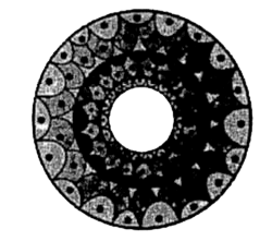
Identify the animalAFasciolaBAscarisCAncylostomaDBoth (a) and (c)The figure given below is a diagrammatic representation of response of organisms to abiotic factors. What do A, B and C represent respectively?
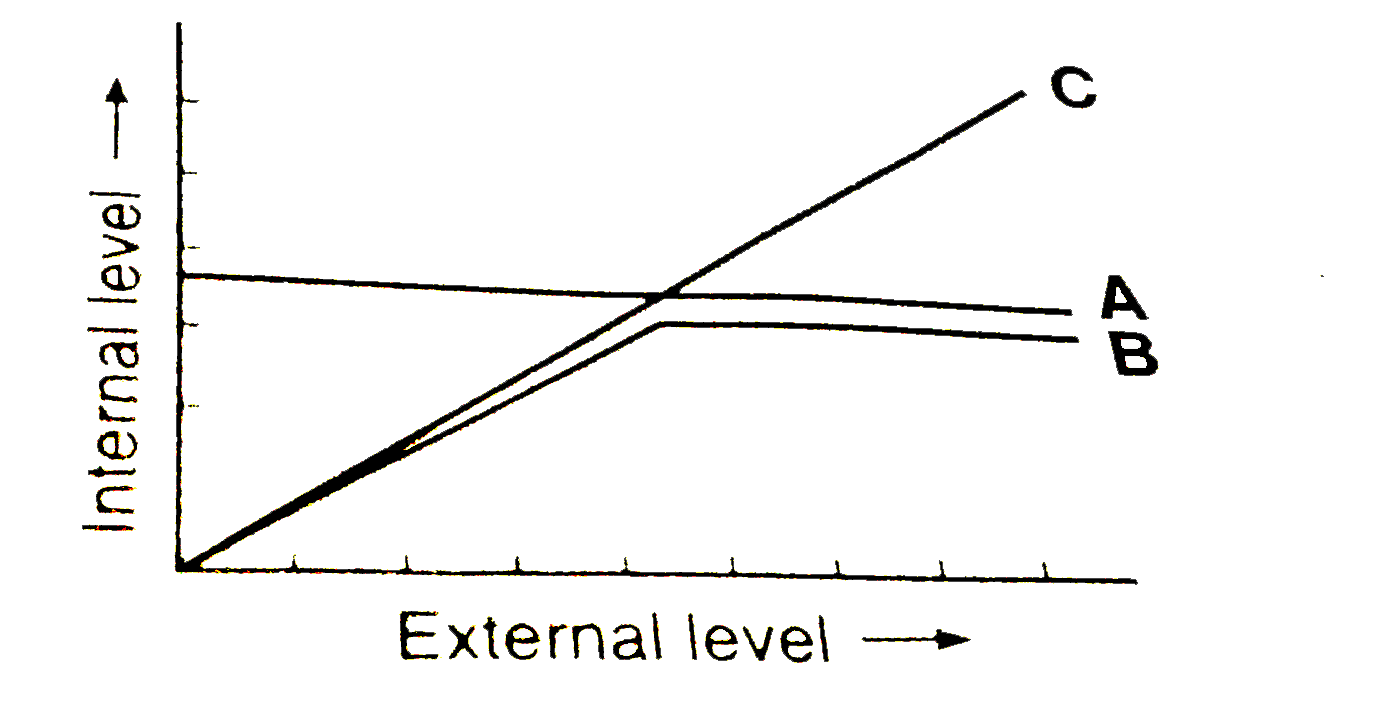 A(A) conformer (B) regulator (C ) partial regulatorB(A) regulator (B) partial regulator (C ) conformerC(A) partial regulator (B) regulator (C ) conformerDregulator (B) conformer (C ) partial regulator
The figure given below is a diagrammatic representation of response of organisms to abiotic factors. What do (i), (ii) and (iii) represent respectively ?
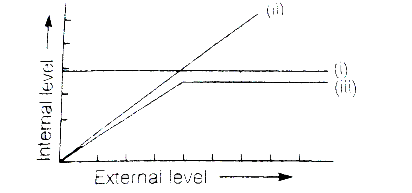 A I II IIIConformerRegulatorPartial regulatorB I II IIIRegulatorPartialConformerregulatorC I II IIIPartial regulatorregulatorConformerD I II IIIRegulatorConformerPartial regulator
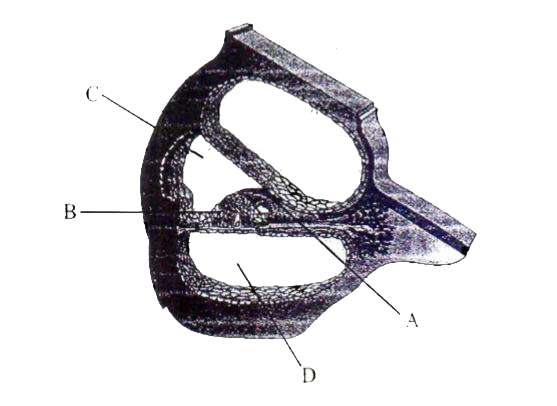
The figure shows a diagrammatic representation of the sectional view of cochlea with labels A, B, C and D. Select the option with correct identification and main characteristic or function:
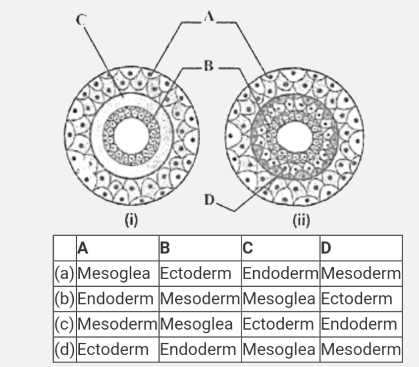
View SolutionIdentify the labelled parts A to D in sections of (i) diploblastic and (ii) triploblastic organisations in animals.
View SolutionDiagrammatically represent the types of chromosomes based on the position of centromere.
View SolutionDiagrammatically represent the experimental steps in cloning and expressing a human gene (say the gene for growth hormone) into a bacterium like E. coli?
View SolutionIn which orientation,a diole placed, in a uniform field is in (i) stable (ii) unstable equilibrioum?
View Solution

