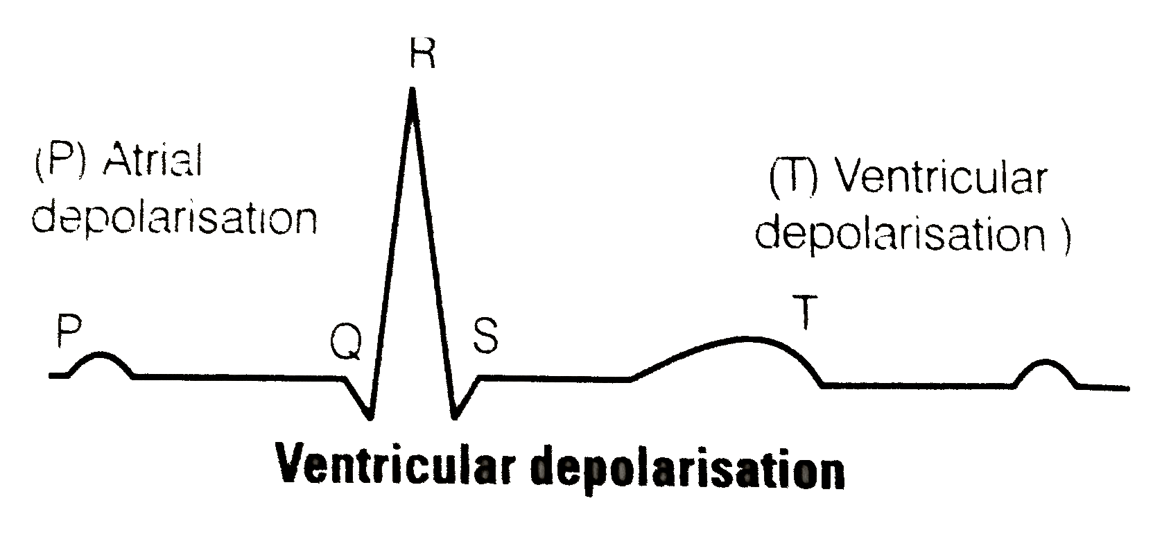
Given below is the diagrammatic representation of a standard ECG.Lable its different peaks.

Video Solution
Text Solution
The representation of a standard ECG diagram
|
Topper's Solved these Questions
- Body Fluids and Circulation BOOK - NCERT EXEMPLARCHAPTER - Body Fluids and Circulation EXERCISE - SHORT ANSWER TYPE QUESTION
9 Videos
- Body Fluids and Circulation BOOK - NCERT EXEMPLARCHAPTER - Body Fluids and Circulation EXERCISE - LONG ANSWER TYPE Q
5 Videos
- Body Fluids and Circulation BOOK - NCERT EXEMPLARCHAPTER - Body Fluids and Circulation EXERCISE - LONG ANSWER TYPE Q
5 Videos
- BIOMOLECULES BOOK - NCERT EXEMPLARCHAPTER - BIOMOLECULES EXERCISE - Long Answer Type Questions
5 Videos
- BREATHING AND EXCHANGE OF GASESBOOK - NCERT EXEMPLARCHAPTER - BREATHING AND EXCHANGE OF GASESEXERCISE - SATQ
7 Videos
Similar Questions
Knowledge Check
The below figure is the diagrammatic representation of standard ECG.
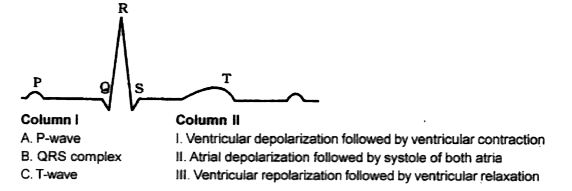 AA - I, B - II, C - IIIBA - III, B - II, C - ICA - II, B - I, C - IIIDA - II, B - III, C - I
Given below is a diagrammatic representation of a glandular tissue
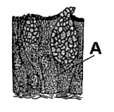
Idientify the structure labelled as 'A' and choose the correct option w.r.t. its its example.AGastric glandBGoblet cellsCOxyntic cellsDSebaceous glandWhich of the following is the diagrammatic representation of standard electrocardiogram (ECG) ?
A B
B C
C D
D
The given diagrammatic representation shows one of the categories of small molecular weight organic compounds in the living tissues. Identify the category shown and the one blank component "X" in it :

View SolutionWhich of the following is the diagrammatic representation of standard electrocardiogram (ECG)?
View SolutionGiven below is a diagrammatic representation of a process taking place in the human body
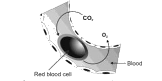
In which of these regions/organs could it be occurring?
i. lungs
ii. Heart
iii. Brain
View SolutionExamine the diagrammatic representation of standard ECG. Select an option with correct matching

View SolutionGiven below is a diagrammatic representation of a body wall of an animal.
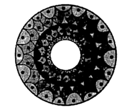
Identify the animal
View Solution

