NEURAL CONTROL AND COORDINATION for Class 11 BIOLOGY
INTRODUCTION
- The functions of the organs/organ systems in our body must be coordinated to maintain homeostasis.
- Coordination is the process through which two or more organs interact and complement the functions of one another. For example, when we do physical exercises, the energy demand is increased for maintaining an increased muscular activity. The supply of oxygen is also increased. The increased supply of oxygen necessitates an increase in the rate of respiration, heartbeat and increased blood flow via blood vessels. When physical exercise is stopped, the activities of nerves, lungs, heart and kidney gradually return to their normal conditions. Thus, the functions of muscles, lungs, heart, blood vessels, kidney and other organs are coordinated while performing physical exercises.
- In our body the neural system and the endocrine system jointly coordinate and integrate all the activities of the organs so that they function in a synchronised fashion.
- The neural system provides an organised network of point-to-point connections for a quick coordination. The endocrine system provides chemical integration through hormones.
NERVOUS TISSUE
- The neural system of all animals is composed of highly specialised cells called neurons which can detect, receive and transmit different kinds of stimuli.The neural organisation is very simple in lower invertebrates.
- For example, in Hydra it is composed of a network of neurons. The neural system is better organised in insects, where a brain is present along with a number of ganglia and neural tissues. The vertebrates have a more developed neural system.
Human Neural System
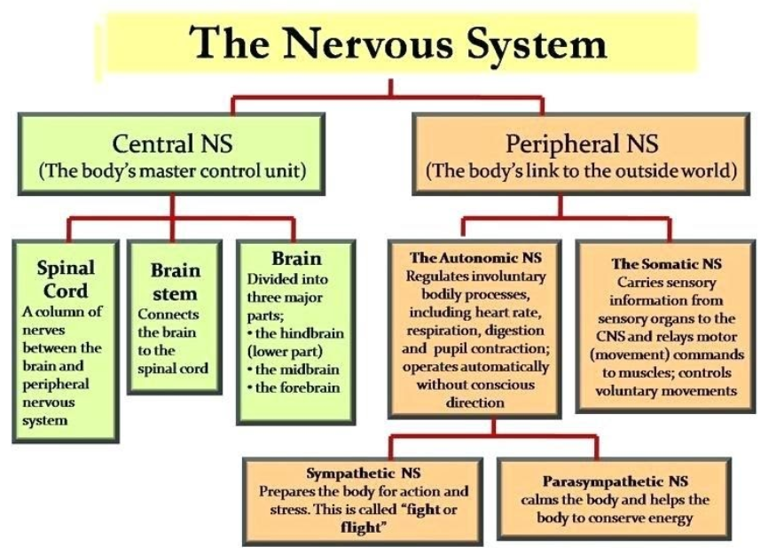
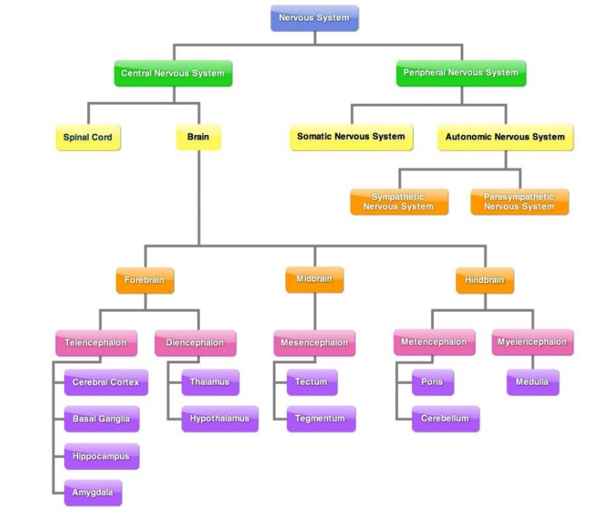
- 4 Major tissue types me se ek nervous tissue hota hai jiske iss chapter me padhenge. Human ke nervous system ko 2 parts me divide kiya gya hai : central nervous system (CNS) and peripheral nervous system (PNS). CNS me brain and spinal cord aati hai and CNS basically processing and control center hai jabki PNS me puri body ki nerves aajati hai. PNS me nerve fibers 2 types ke hote hai : ek body ke different organs se sensory information ko CNS tak lekr jaye, jinko hum afferent fibers kehte hai wo and dusre aise fibers hote hai jo CNS se baki body parts tak information lekr jaye, jinko hum efferent fibers kehte hai. PNS ko hi further Somatic neural system and autonomic neural system me divide kiya jata hai. Somatic neural system ka kaam hai CNS se skeletal muscles tak information lekr jana, jo ki voluntary tissue hai. Autonomic nervous system me wo fibers hote hai jo CNS se involuntary organs and smooth muscles tak information lekar jate hai. Issi autonomic system ko further sympathetic neural system and parasympathetic neural system me divide kiya gya hai, and yeah dono ek dusre ke opposite kaam karte hai yani ki antagonistic hai. For example, neurons of the sympathetic nervous system increase heart rate, and neurons of the parasympathetic nervous system slow it down. In general, the parasympathetic nervous system takes care of “rest-and-digest” activities, and the sympathetic nervous system helps support exercise or emergency actions—the so-called “fight-or-flight” responses.
- Nervous tissue me 2 types ke cells hote hai : neurons and neuroglia. Neurons basically neural system ki basic functional unit hai jo nerve impulses yani ki information ko propagate karte rehte hai. Yeah cells itne specialized hai ki inki mitotic division ki ability bhi khatam ho chuki hai.
- Neuroglia chotey cells hai but inka number, neurons se bahut jyada hai. Inka kaam hai neurons ko support, nourish, protect karna and interstitial fluid ko maintain karna. Yeah cells divide kar sakte hai.
- Neurons, electrical stimulus ke respect me excite hone wale cells hai.
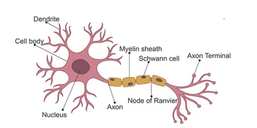
- Mostly neurons ke 3 parts hote hai : cell body, dendrites and an axon.
- Cell body ko hi perikaryon bhi bol dete hai, isme nucleus and cytoplasm hota hai along with other cell organelles. Free ribosomes and RER ke clusters kafi prominent dikhte hai and inhi ko hum nissl bodies ya nissl granules keh dete hai.
- cell bodies ka cluster agar CNS me hai toh nucleus kehlata hai and agar PNS me hai toh ganglion kehlata hai.
- Ek cell body se bahut sari processes/outgrowths nikalti hai jinko dendrites bol diya jata hai. Inhi dendrites ke through ek neuron sensory information leta hai. Dendrites ki plasma membrane pe bahut sare receptors hote hai.
- Axon basically dendrite ke comparison me kafi thick outgrowth hai jo ki dendrites ki tarah highly branched nahi hoti lekin axon ke right angle pe kuch side branches nikalti hai jinko axon collaterals bola jata hai. Soma ke jis part se axon shuru hoti hai usko axon hillock bola jata hai. Axon me RER nahi hota. Axon ke cytoplasm ko hi axoplasm bola jata hai and iski plasma membrane ko axolemma bolte hai. Axon end me jakr bahut sari fine process me divide karti hai and each process ends into a knob like structure known as axon terminal. Yeah axon terminal next neuron ke dendrites ke sath contact establish karte hai lekin directly touch nahi karte, in dono ke beech me thoda sa gap hota hai jo ki synapse kehlata hai.
- Multipolar neurons : bahut sarey dendrites and ek axon inki pehchaan hai. CNS me most of the neurons, multipolar hi hai.
- Bipolar neurons : ek dendrite and ek axon inki pehchaan hai. Ex : retina of eye, inner ear and brain ke olfactory area me aise neurons milte hai.
- Pseudounipolar neurons : inki cell body se ek process nikalti hai jo ki thodi se dur jakr 2 me divide kar jati hai, ek se further kuch distance pe dendrites emerge karte hai jabki dusri se axon terminals. Cell body se directly koi dendrites nahi aate. Yeah neurons mostly sensory receptors ki tarah function karte hai.
- Afferent / sensory neurons : yeah sensory organs se CNS tak information convey karte hai and mostly yeah unipolar hote hai nature me.
- Motor neuron ya efferent neurons : yeah CNS se effector organs tak information lekr aate hai. Generally multipolar nature ke hote hai.
- Interneurons : inhe association neurons bhi bola jata hai. Yeah CNS me hi sensory and motor neurons ke beech located hai. Yeah sensory neurons se aane wali sensory information ko process karke ek motor response generate karte hai jo ki motor neurons carry karte hai.
- Yeah cells glue ka kaam karte hai in the sense ki neurons ko hold karke rakhte hai. CNS and PNS me alag alag neuroglia cells hote hai.
- Neuroglia of CNS : astrocytes (star shaped cells), oligodendrocytes (CNS me myelin sheath bnate hai), Microglia (phagocytic cells), Ependymal cells (secrete cerebrospinal fluid).
- Neuroglia of PNS : Schwann cells (form myelin sheath around axons in PNS), Satellite cells (regulate the exchanges of materials between neuronal cell bodies and interstitial fluid).
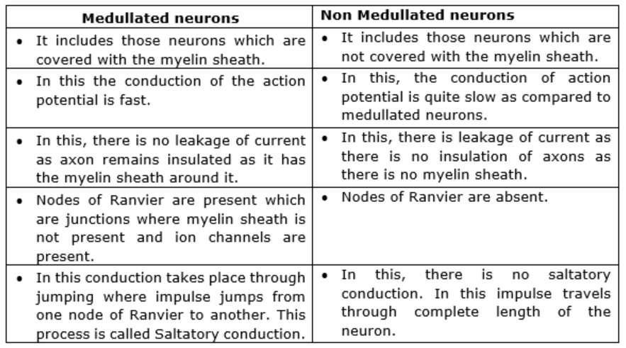
- Myelination : myelin sheath is a multilayered lipid and protein covering around some axons that insulates them and increases the speed of nerve impulse conduction. Jin neurons ke around myelin sheath hoti hai unlo myelinated neurons kehte hai and jinke around nahi hoti unko hum unmyelinated neurons kehte hai. Myelin sheath, axon ki longitudinal axis ke continuation me start se end tak nahi hoti. Yani ki beech beech me axon ka chota sa hissa asia hota hai jhan myelin sheath nahi hoti, un hisso ki hum nodes of ranvier kehte hai. Unmyelinated nerve fibre is enclosed by a Schwann cell that does not form a myelin sheath around the axon, and is commonly found in autonomous and the somatic neural systems.
- PNS me Axons ke bundles ko hi nerve bola jata hai jabki CNS me axons ke bundle ko tract bola jata hai.
- White matter is composed primarily of myelinated axons. The whitish color of myelin gives white matter its name. The gray matter of the nervous system contains neuronal cell bodies, dendrites, unmyelinated axons, axon terminals, and neuroglia. It appears grayish, rather than white, because the Nissl bodies impart a gray color and there is little or no myelin in these areas. Spinal cord ki core me gray matter (shaped like a butterfly or the letter H in transverse section ) hota hai jabki outer side me white matter hota hai. Brain (cerebrum & cerebellum) me surface par gray matter hota hai jabki uske neeche white matter hota hai.
- Neurons me positively and negatively charged ions ke movement and distribution ke karan hi ek potential generate hota hai jiske action potential ya nerve impulse kehte hai. Toh iss electrical activity ka karan hai neuron me paye jane wale different ion channels jo membrane ki +ve and -ve charge ke prati permeability ko regulate karte hai.
- Leak channels basically open state me rehte hai toh inke through ions nikalti rehti hai. Har ion ka apna alag channel hota hai.
- Ligand gated channels aise channels hai jo kisi chemical entity ke response me open ya close honge. Yani ki koi chemical aagya toh yeah open ho jayenge and inke through ions flow karenge otherwise nahi.
- Mechanically gated channels basically mechanical stimulus ke response me open and close hote hai.
- Voltage gated channels, voltage me change ko sense karke open and close hote hai.
Histology of Nervous Tissue
Neurons
Structural Classification of Neurons
Functional CLassification of Neurons
Neuroglia
Gray & White matter
Ion Channels in Neurons
NEURONS
- These are structural and functional units of the neural system. A neuron is a microscopic structure composed of three major parts, namely, the cell body, dendrites and axon.
- The cell body contains cytoplasm with typical cell organelles, spherical nucleus and certain granular bodies called Nissl’s granules and neurofibrils.Nissl’s granules are masses of ribosomes and rough endoplasmic reticulum. They synthesise proteins in the cell.
- Process of neuron(neurites):
- Short fibres which branch repeatedly and project out of the cell body also contain Nissl’s granules and are called dendrites. These fibres transmit impulses towards the cell body.
- The axon/axis cylinder(neuraxon) is a long fibre, the distal end of which is branched. Each branch terminates as a bulb-like structure called synaptic knob which possess synaptic vesicles containing chemicals called neurotransmitters. The axons transmit nerve impulses away from the cell body to a synapse or to a neuro-muscular junction.
- The part of the cyton from where the axon arises is called axon hillock. It is most sensitive part of the neuron. The axon ends in a group of branches, the terminal arborizations.
- Certain axons also give rise to side branches, called collateral fibres.
- Nissl’s granules, Golgi bodies, ribosomes and fat globules are absent in axoplasm
- .The plasma membrane and neuroplasm of axon are respectively called axolemma and axoplasm.
TYPES OF NEURONS
- On the basis of number of axon and dendrites, the neurons are divided into three types, i.e.
- Unipolar neurons - These have a single process arising from cyton. Eg early embryos of invertebrates and vertebrates.
- Bipolar neurons - These neurons have only two processes, an axon at one end and a dendrite at another end. EG. retina of the eyes, olfactory epithelium and cochlear and vestibule ganglia.
- Multipolar neuron - These neurons have several dendrites and one axon. Eg cerebral cortex.
- On the basis of functions, neurons are of following three types,i.e.
- Sensory(receptor or afferent) neurons - These connect sense organs with CNS.
- Motor(effector or efferent) neurons - These connect the CNS to the effectors.
- Interneurons(connector, relaying or adjustor neurons) - These are present in the CNS and occur between the sensory and motor neurons for distant transmission of impulses.
Medullary sheath
- It is composed of substances called myelin, which contain lipids, proteins and water.It serves as an insulating layer, preventing loss of energy of the nerve impulse during its passage along the fibre.
- It is absent at certain points called nodes of Ranvier. The part of a nerve fibre between two successive nodes of ranvier is called internode. Each segment of the myelin sheath is formed by one Schwann cell.
Neurilemma
- Outside the myelin sheath there is a layer of Schwann cell which is called neurilemma, which is continuous over the nodes of Ranvier.
- A nerve consists of several bundles of nerve fibres.
Neuroglia
- Neuroglial cells are specialised cells found in the brain and spinal cord supporting the neurons and their fibres.
- These comprise about 50% of all the brain cells.
Ependymal cells
- These are present in ventricles and the central canal of the spinal cord.
- Their shape varies from squamous to columnar and free surfaces of the cells bear numerous microvilli and cilia, which help in the absorption and flow of cerebrospinal fluid respectively.
Neurosecretory cells
- These specialised nerve cells function as endocrine cells.
- Neurosecretory cells of the hypothalamus of the vertebrate brain secrete neurohormones.
GENERATION AND CONDUCTION OF NERVE IMPULSE
- Nerve impulse - A wave of reversed polarity or depolarisation(action potential) moving down an axon is called a nerve impulse.
- When a neuron is suitably stimulated, an electrical disturbance is generated which swiftly travels along its plasma membrane. Arrival of the disturbance at the neuron’s endings or output zone, triggers events that may cause stimulation or inhibition of adjacent neurons and other cells.
- Neurons are excitable cells because their membranes are in a polarised state.
- Most accepted mechanism of nerve impulse conduction is ionic theory proposed by Hodgkin and Huxley. This theory states that nerve impulse is an electro-chemical process governed by differential permeability of axolemma to sodium and potassium ions which in turn is regulated by the electric field.
- Different types of ion channels are present on the neural membrane. These ion channels are selectively permeable to different ions.
POLARISATION
- When a neuron is not conducting any impulse, i.e resting, the axonal membrane is comparatively more permeable to potassium ions and nearly impermeable to sodium ions. Similarly, the membrane is impermeable to negatively charged proteins present in the axoplasm.
- Consequently, the axoplasm inside the axon contains high concentration of K+ and negatively charged proteins and low concentration of Na+.
- In contrast the fluid outside the axon contains a low concentration of K+, a high concentration of Na+ and thus forms a concentration gradient.
- These ionic gradients across the resting membrane are maintained by the active transport of ions by the sodium-potassium pump which transport 3 Na+ outwards for 2K+ into the cell.
- As a result, the outer surface of the axonal membrane possesses a positive charge while its inner surface becomes negatively charged and therefore is polarised.
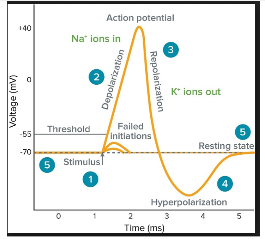
- The electrical potential difference across the resting plasma membrane is called the resting potential (-70mv.)
Nerve Impulse : Generation & Conduction
- Resting membrane potential : Neurons me resting stage me axoplasm me negatively charge ions and proteins ke hone ke karan, axon ki membrane ki inner side pe negative charge accumulate hua hota hai and neurons ke bahar fluid me positively charge sodium ions ke karan bahar +ve charge accumulate hota hai. Axon ki membrane ke across yahi +ve and -ve charge ki differential distribution ke karan ek potential develop hota hai jiske hum resting membrane potential (RMP) kehte hai. RMP ki general value hoti hai = -70mV. Minus sign basically yeah indicate kar rha hai ki neuron ki inside, bahar ke environment ke comparison me negative hai.
- Issi +ve and -ve charge ke differential distribution ke karan neuron ki inner surface -ve pole and outer membrane +ve pole ki tarah behave karti hai. Toh iss polarity ke develop hone ke karan hi resting stage me bhi ek neuron polarized hota hai.
- Three factors that contribute to the resting membrane potential :
- Because the plasma membrane has more K+ leak channels (blue) than Na+ leak channels, the number of K+ ions that leave the cell is greater than the number of Na+ ions that enter the cell. As more and more K+ ions leave the cell, the inside of the membrane becomes increasingly negative and the outside of the membrane becomes increasingly positive.
- Trapped anions cannot follow K+ out of the cell because they are attached to non diffusible molecules such as ATP and large proteins.
- The electrogenic Na+–K+ ATPase expels 3 Na+ ions for every 2 K+ ions imported.
- Ab jab bhi koi stimulus apply karte hai toh axolemma ke jis hisse pe stimulus lagta hai, depending upon the type of stimulus (ki chemical stimulus hai ya mechanical stimulus hai ya electrical stimulus hai), respective sodium channels open hojate hai jinke karan axoplasm me +vely charge sodium ions flow karna shuru kar deti hai. Ab axolemma ki inner side bahar ke comparison me jyada positively charged hai and outer side of membrane is negatively charged. Yani ki resting stage ke comparison me ab jo polarity thi wo reverse ho gyi. Yani ki membrane ab depolarise ho chuki hai jiske hum nerve impulse generation kehte hai.
- Ab axon ki jis site me nerve impulse generate hua hai uske immediately next part me abhi bhi normal resting stage conditions hai yani ki inner side is -vely charged and outer surface is positively charged. Toh electric current is site par flow karega and similarly aise karte karte axons ke terminals tak yeah electric current yani ki nerve impulse flow karegi. Issi ko hum nerve impulse conduction kehte hai.
- Ab jaise ki sodium ions ander aaie and membrane positively charged hui and nerve impulse generate and conduct hui, sath ki membrane potassium ions ke liye permeable ho jati hai and potassium ions yani ki +ve charge axoplasm se bahar move karna shuru kar deta hai and it restores the normal polarity, yani ki jitna positive charge ander aaya tha utna bahar gya and membrane firse resting stage polarity par aagyi. Iss phase ko hum repolarisation kehte hai. Kafi normally yeah bhi dekha gya hai ki repolarisation ke waqt jo potassium channels thy wo jyada time tak khule rehte hai toh normal se jyada positive charge bahar nikalne ke karna membrane ki inner side resting stage ke comparison me jyada negative ho jati hai and isko hum hyperpolarisation kehte hai.
- Yeah bahut hi basic overview tha nerve impulse generation and conduction ka. There are a lot of factors which we haven’t yet touched because they are not a part of CBSE syllabus. We will deal with them in higher classes.
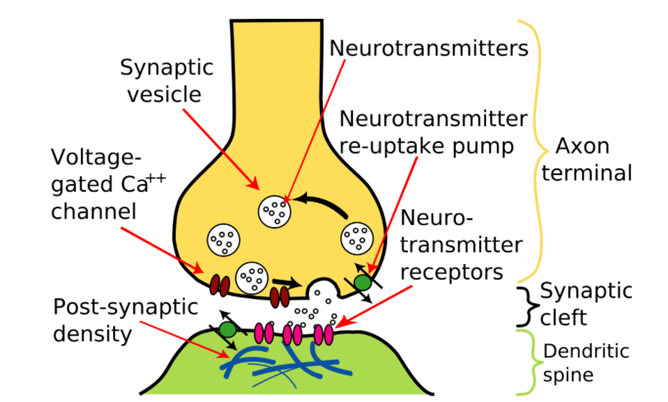
- Ab jo yeah impulse generate hui, then axon ki length ke alon nerve terminals tak conduct hui, yeah finally axon terminals tak jab pahcuti hai toh axon terminals me kuch chemical messengers vesicles me packed hote hai. Inn vesicles ko hum synaptic vesicles kehte hai kyunki nerve impulse ke yhaan tak aane ke baad yeah yeah vesicles axon terminal ki membrane ke sath fuse karke, exocytosis ki process ke dwara, apne contents (neurotransmitters) ko synaptic cleft me release kardeti hai.
- Synaptic cleft se yeah chemical next neuron ki dendrite par located receptors tak jakr unse bind karte hai and next neuron me new impulse ko generate karte hai.
- Jis neuron se impulse aa rahi hai and jis neuron ne neurotransmitters ko synaptic cleft me release kia hai usko hum pre-synaptic neuron kehte hai and jis neuron tak synapse se neurotransmitters travel karke jate hai and bind karke new impulse generate karte hai usko hum post synaptic neuron kehte hai.
DEPOLARISATION
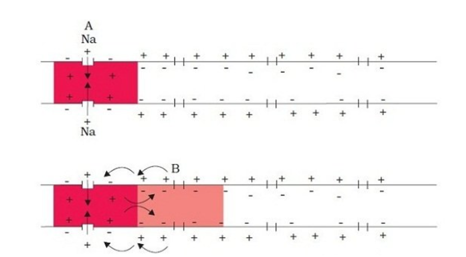
- When a stimulus is applied at a site, point A on the polarised membrane, the membrane at the site A becomes freely permeable to Na+.
- This leads to a rapid influx of Na+ followed by the reversal of the polarity at that site, i.e., the outer surface of the membrane becomes negatively charged and the inner side becomes positively charged.
- The polarity of the membrane at the site A is thus reversed and hence depolarised. The electrical potential difference across the plasma membrane at site A is called the action potential(+20,-40mv), which is in fact termed as a nerve impulse(spike potential).
- At sites immediately ahead, the axon (e.g., site B) membrane has a positive charge on the outer surface and a negative charge on its inner surface. As a result, a current flows on the inner surface from site A to site B. On the outer surface current flows from site B to site A to complete the circuit of current flow.
- Hence, the polarity at the site is reversed, and an action potential is generated at site B. Thus, the impulse (action potential) generated at site A arrives at site B.
- The sequence is repeated along the length of the axon and consequently the impulse is conducted.
POLARISATION
- When a neuron is not conducting any impulse, i.e resting, the axonal membrane is comparatively more permeable to potassium ions and nearly impermeable to sodium ions. Similarly, the membrane is impermeable to negatively charged proteins present in the axoplasm.
- Consequently, the axoplasm inside the axon contains high concentration of K+ and negatively charged proteins and low concentration of Na+.
- In contrast the fluid outside the axon contains a low concentration of K+, a high concentration of Na+ and thus forms a concentration gradient.
- These ionic gradients across the resting membrane are maintained by the active transport of ions by the sodium-potassium pump which transport 3 Na+ outwards for 2K+ into the cell.
- As a result, the outer surface of the axonal membrane possesses a positive charge while its inner surface becomes negatively charged and therefore is polarised.
- The electrical potential difference across the resting plasma membrane is called the resting potential (-70mv.)
REPOLARISATION
- The rise in the stimulus-induced permeability to Na+ is extremely short lived.
- It is quickly followed by a rise in permeability to K+ .
- Within a fraction of a second, K+ diffuses outside the membrane and restores the resting potential of the membrane at the site of excitation and the fibre becomes once more responsive to further stimulation(Repolarisation).
- The whole process of depolarisation and repolarisation is very fast and takes about 1 to 5ms.
- The last movement of ions is thought to take place by an active transport mechanism called sodium potassium pump.
- During repolarization as the cell returns to resting potential, it is ready to receive another stimulus.
- At the end of action potential most neurons undergo a refractory period in which the membrane potential briefly becomes even more negative than it normally is at rest, It is called hyperpolarizing phase.
- This occurs because of increased potassium permeability that exists because voltage gated potassium channels remain open for longer duration. It results in transmission of nerve impulse only in forward direction.
DEPOLARISATION
- When a stimulus is applied at a site, point A on the polarised membrane, the membrane at the site A becomes freely permeable to Na+.
- This leads to a rapid influx of Na+ followed by the reversal of the polarity at that site, i.e., the outer surface of the membrane becomes negatively charged and the inner side becomes positively charged.
- The polarity of the membrane at the site A is thus reversed and hence depolarised. The electrical potential difference across the plasma membrane at site A is called the action potential(+20,-40mv), which is in fact termed as a nerve impulse(spike potential).
- At sites immediately ahead, the axon (e.g., site B) membrane has a positive charge on the outer surface and a negative charge on its inner surface. As a result, a current flows on the inner surface from site A to site B. On the outer surface current flows from site B to site A to complete the circuit of current flow.
- Hence, the polarity at the site is reversed, and an action potential is generated at site B. Thus, the impulse (action potential) generated at site A arrives at site B.
- The sequence is repeated along the length of the axon and consequently the impulse is conducted.

SYNAPSE
- Sherrington first used the term synapse.
- A nerve impulse is transmitted from one neuron to another through junctions called synapse. A synapse is formed by the membranes of a presynaptic neuron and a postsynaptic neuron, which may or may not be separated by a gap called synaptic cleft.
- Chemical synapse:
- At a chemical synapse, the membranes of the pre- and postsynaptic neurons are separated by a fluid-filled space called synaptic cleft.
- Chemicals called neurotransmitters are involved in the transmission of impulses at these synapses. The axon terminals contain vesicles filled with these neurotransmitters.When an impulse (action potential) arrives at the axon terminal, it stimulates the movement of the synaptic vesicles towards the membrane where they fuse with the plasma membrane and release their neurotransmitters in the synaptic cleft.
- The released neurotransmitters bind to their specific receptors, present on the postsynaptic membrane.
- This binding opens ion channels allowing the entry of ions which can generate a new potential in the postsynaptic neuron. The new potential developed may be either excitatory or inhibitory.
- Electrical synapse:
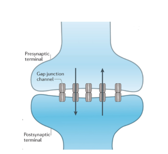
- At electrical synapses, the membranes of pre- and postsynaptic neurons are in very close proximity.
- Electrical current can flow directly from one neuron into the other across these synapses.
- Transmission of an impulse across electrical synapses is very similar to impulse conduction along a single axon.
- Impulse transmission across an electrical synapse is always faster than that across a chemical synapse.
- Electrical synapses are rare in our system
- Synapse, A one-way valve : The synapse cannot transmit an impulse in the reverse direction as dendrites cannot secrete neurotransmitters. Thus, the synapse acts a one-way valve, allowing the conduction of impulse from axon to dendrite only.
- Synaptic delay: Transmission of an impulse across a synapse is slower than its conduction along a neuron . This is because of the time needed for the release of a neurotransmitter. Its diffusion through the synaptic cleft, and its action on the postsynaptic membrane.
TYPES OF SYNAPSE
- There are two major types of synapse:
PROPERTIES OF SYNAPSE
REPOLARISATION
- The rise in the stimulus-induced permeability to Na+ is extremely short lived.
- It is quickly followed by a rise in permeability to K+ .
- Within a fraction of a second, K+ diffuses outside the membrane and restores the resting potential of the membrane at the site of excitation and the fibre becomes once more responsive to further stimulation(Repolarisation).
- The whole process of depolarisation and repolarisation is very fast and takes about 1 to 5ms.
- The last movement of ions is thought to take place by an active transport mechanism called sodium potassium pump.
- During repolarization as the cell returns to resting potential, it is ready to receive another stimulus.
- At the end of action potential most neurons undergo a refractory period in which the membrane potential briefly becomes even more negative than it normally is at rest, It is called hyperpolarizing phase.
- This occurs because of increased potassium permeability that exists because voltage gated potassium channels remain open for longer duration. It results in transmission of nerve impulse only in forward direction.
PHYSICAL PROPERTIES OF NEURONS
- All or none law: The stimulation is conducted only when the threshold is broken. If the stimulation is too weak to break the axon’s threshold, impulse is not established, but if the intensity of stimulation is much more than the threshold value, impulse conduction remains normal. Thus, the action potential obeys this all or none law.
- Saltatory conduction: Myelin sheath is lipid rich region of high electrical resistance hence action potential jumps as an electric current from one node to next. The arrival of the current at that node opens its Na+ channels creating another current that passes on the next node and so on. This very fast form of nerve impulse conduction is known as saltatory conduction.
- Conduction speed: According to the nature of neuron transmission speed varies from 0.5ms-1 to over 100ms-1. Two factors are important in determining the speed of conduction.
- The diameter of the axon - The greater is the diameter the faster is the speed of transmission.
- The myelin sheath.
SYNAPSE
- Sherrington first used the term synapse.
- A nerve impulse is transmitted from one neuron to another through junctions called synapse. A synapse is formed by the membranes of a presynaptic neuron and a postsynaptic neuron, which may or may not be separated by a gap called synaptic cleft.
- Chemical synapse:
- At a chemical synapse, the membranes of the pre- and postsynaptic neurons are separated by a fluid-filled space called synaptic cleft.
- Chemicals called neurotransmitters are involved in the transmission of impulses at these synapses. The axon terminals contain vesicles filled with these neurotransmitters.When an impulse (action potential) arrives at the axon terminal, it stimulates the movement of the synaptic vesicles towards the membrane where they fuse with the plasma membrane and release their neurotransmitters in the synaptic cleft.
- The released neurotransmitters bind to their specific receptors, present on the postsynaptic membrane.
- This binding opens ion channels allowing the entry of ions which can generate a new potential in the postsynaptic neuron. The new potential developed may be either excitatory or inhibitory.
- Electrical synapse:
- At electrical synapses, the membranes of pre- and postsynaptic neurons are in very close proximity.
- Electrical current can flow directly from one neuron into the other across these synapses.
- Transmission of an impulse across electrical synapses is very similar to impulse conduction along a single axon.
- Impulse transmission across an electrical synapse is always faster than that across a chemical synapse.
- Electrical synapses are rare in our system
- Synapse, A one-way valve : The synapse cannot transmit an impulse in the reverse direction as dendrites cannot secrete neurotransmitters. Thus, the synapse acts a one-way valve, allowing the conduction of impulse from axon to dendrite only.
- Synaptic delay: Transmission of an impulse across a synapse is slower than its conduction along a neuron . This is because of the time needed for the release of a neurotransmitter. Its diffusion through the synaptic cleft, and its action on the postsynaptic membrane.
TYPES OF SYNAPSE
-There are two major types of synapse:PROPERTIES OF SYNAPSE
NERVOUS SYSTEM
PHYSICAL PROPERTIES OF NEURONS
- All or none law: The stimulation is conducted only when the threshold is broken. If the stimulation is too weak to break the axon’s threshold, impulse is not established, but if the intensity of stimulation is much more than the threshold value, impulse conduction remains normal. Thus, the action potential obeys this all or none law.
- Saltatory conduction: Myelin sheath is lipid rich region of high electrical resistance hence action potential jumps as an electric current from one node to next. The arrival of the current at that node opens its Na+ channels creating another current that passes on the next node and so on. This very fast form of nerve impulse conduction is known as saltatory conduction.
- Conduction speed: According to the nature of neuron transmission speed varies from 0.5ms-1 to over 100ms-1. Two factors are important in determining the speed of conduction.
- The diameter of the axon - The greater is the diameter the faster is the speed of transmission.
- The myelin sheath.
BRAIN
- The brain is the central information processing organ of our body, and acts as the ‘command and control system’.
- It controls the voluntary movements, balance of the body, functioning of vital involuntary organs (e.g., lungs, heart, kidneys, etc.), thermoregulation, hunger and thirst, circadian (24-hour) rhythms of our body, activities of several endocrine glands and human behaviour. It is also the site for processing of vision, hearing, speech, memory, intelligence, emotions and thoughts.
- Brain contains about 100 billion neurons. Number of neuroglial cells present in the brain is one trillion.
Central Nervous System
- CNS me brain and spinal cord aate hai. Pehle baat karenge brain ki.
Brain
- Brain skull me located hai. Brain ke aroun 3 protective layers hoti hai jinko hum meninges kehte hai. Outside to inside they are : dura mater, arachnoid and pia mater. Pater mater directly nervous tissue ke contact me hoti hai. Yhi protective layers spinal cord me bhi hoti hai but spinal cord ki dura mater single layered hoti hai jabki brain ki double layered.
- Nervous tissue ectodermally derived tissue hai jhaan embryo me organogenesis ke waqt ectoderm layer se neural tube banti hai. Then neural tube se 3 primary brain vesicles banti hai jinko hum prosencephalon, mesencephalon, and rhombencephalon kehte hai.
- Fir secondary brain vesicles banti hai because of further subdivisions in prosencephalon and rhombencephalon. Prosencephalon, or forebrain, gives rise to the telencephalon and diencephalon, and the rhombencephalon, or hindbrain, develops into the metencephalon and myelencephalon.
- Telencephalon se lateral ventricles and cerebrum develop hota hai.
- Diencephalon se thalamus, hypothalamus, epithalamus and 3rd ventricle develop hota hai.
- Mesencephalon se midbrain develop hota hao.
- Metencephalon se pons, cerebellum and 4th ventricle ka upper part develop hota hai.
- Myelencephalon se medulla oblongata and 4th ventricle ka lower part bant ahai.
- Agar uper wala samjh nhi aaya toh bho koi baat nhi. Jaise jaise aagey discuss karenge things will make much more sense. Brain ke normally 3 part hote hai : forebrain, midbrain and hind brain. Hind brain se spinal cord originate karti hai : yeah toh yaad rakhna hai.
- Brain me kuch cavities hoti hai jinme cerebrospinal naam ki fluid hoti hai. Inhi cavities ko hum ventricles keh dete hai. Aise 4 ventricles brian me hote hai. 2 lateral ventricles hote hai kyunki brain ke lateral aspect me present hai. Lateral ventricles forebrain ke dono hemispheres me present hote hai. The third ventricle is a narrow, slitlike cavity along the midline superior (superior ka matlab hai “uper ki taraf”) to the hypothalamus and between the right and left halves of the thalamus. The fourth ventricle lies between the brainstem and the cerebellum. Inhi ventricles ki roof pe ependymal cells hote hai jo CSF secrete karte hai. CSF buoyancy provide karti hai and nutrition provide karegi.
- Forebrain me cerebrum, thalamus and hypothalamus naam ke structures aate hai. Cerebrum brain ka major part hai and yeah right & left hemispheres me divided hota hai.
- Nerve fibres ke tract jinko corpus callosum bola jata hai, wo in dono hemispheres ko apas me connect karte hai. Outer side grey matter hota hai jabki inner side white matter hota hai.
- Cortex plain nahi hota rather highly convoluted surface hai jisme bahut sare folds hote hai.
- Cerebral cortex me 3 main areas hote hai : sensory areas jo sensory information ko recieve karte hai, motor areas jo ki motor output ke liye responsible honge and association areas jo ki na toh totally sensory hai and na hi totally motor areas hai. Yeah association areas basically complex functions like intersensory associations, memory and Communication ke liye responsible hote hai.
- Thalamus paired oval structures hai jo ki roughly 3cm ke hote hai. The thalamus is the major relay station for most sensory impulses that reach the primary sensory areas of the cerebral cortex from the spinal cord and brainstem (Yani sensory information pehle thalamus me aati hai and then thalamus decide karta hai ki brain ke kis hisse me iss information ko bhejna hai). In addition, the thalamus contributes to motor functions by transmitting information from the cerebellum and basal nuclei to the primary motor area of the cerebral cortex. The thalamus also relays nerve impulses between different areas of the cerebrum and plays a role in the maintenance of consciousness.
- Hypothalamus : thamus ke neeche hi hypothalamus located hota hai. Yeah homeostasis ko main karta hai jaise ki temperature, blood glucose level, osmotic concentration etc. Hypothalamus is also a major regulator of visceral activities, like regulation of heart rate, movement of food through the gastrointestinal tract, and contraction of the urinary bladder. Hypothalamus bahut sare hormones bhi produce karta hai jinko hum next chapter me study karenge. Eating, drinking, emotional & behavioural patterns (rage, aggression, pain, and pleasure, and the behavioral patterns related to sexual arousal etc. ), circadian rhythms etc bhi hypothalamus hi regulate karta hai. Amygdala and hippocampus bhi hypothalamus ke sath sexual behaviour and memomory formation me participate karte hai.
- Forebrain ke thalamus/hypothalamus and hind brain ke pon ke beech ka hissa midbrain kehlata hai. Midbrain, hindbrain ke sath milkar brain stem kehlata hai. Midbrain ki dorsal side par 4 swellings hoti hai jinko corpora quadrigemina kaha jata hai. Uper ki 2 superior colliculi kehlati hai jo ki vision me role play kerti hai jabki lower 2 swellings ko inferior colliculi bola jata hai and yeah hearing me kaam aati hai.
- Hindbrain me pons, cerebellum (little brain) and medulla oblongata hoti hai. Pons ke fibers brain ki different regions ko interconnect karti hai and issi me pontine respiratory group bhi hota hai. Medulla me respiratory, cardiovascular reflexes and gastric secretions ko regulate karne ke liye respective centers hote hai sath hi limbs and trunk ki voluntary movements, vomiting center, deglutition center, sneezing, coughing, hiccuping jaisi activities bhi medulla ke hi control me hoti hai.
- Kisi stimulus ke prati aisa involuntary action jisme conscious effort na lage wo reflex arc kehlata hai.
- Most reflex arcs jaise ki jyada garam object ko touch karne pe haath peeche htana ya kante pe touch hote hi leg retract kar lena jaise kaam spinal cord bina brian ko involve kiye apne level pe handle karleta hai. Inko hum reflex arcs bolte hai.
- Reflex arc me involved neurons ke through information travel karte hue jo path follow karegi usko reflex pathway bola jata hai.
- Slamming on the brakes in an emergency is another example of reflex action.
- Most reflexes jo spinal level pe operate hote hai unko hum spinal reflexes bolte hai jabki jin reflexes me brainstem involve hojati hai unko hum cranial reflexes bolte hai for example, the tracking movements of your eyes as you read this sentence. Matlab sentence padhte hue apne yeah nhi socha na ki ab eye left to right ya right to left move karni padengi, soch kar toh aisa karte nahi; bas apne aap ho jata hai toh yeah ek cranial reflex hai.
- Again there are multiple reflexes like autonomic reflexes, flexor and crossed extensor reflex vgarah and with each topic the complexity level increase but in sabki jarurat nahi hai - na CBSE me and na hi competitive exams me.
Forebrain
Midbrain
Hindbrain
Reflex action & Reflex arc
MENINGES
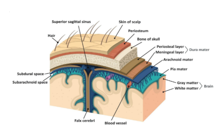
- They are protective membranous coverings of the brain and spinal cord. Meninges are three in number. They are dura mater, arachnoid and pia mater.
- Dura Mater- It is an outer thick layer of fibrous tissue that lines the cranial cavity. Dura mater resists the movement of the brain inside the cranium so that stretching and breaking of blood vessels do not occur.
- Arachnoid - It is a middle meninx, which is thin, webby, porous.
- Subdural space between the dura mater and arachnoid is filled with fluid which is not cerebrospinal fluid.
- Pia Mater - It is moderately tough inner meninx which is highly vascularised and pigmented.
- The space between the arachnoid and pia mater is called subarachnoid space. It contains cerebrospinal spinal fluid and connective tissue strands.
NERVOUS SYSTEM

COMPONENTS OF BRAIN
BRAIN
- The brain is the central information processing organ of our body, and acts as the ‘command and control system’.
- It controls the voluntary movements, balance of the body, functioning of vital involuntary organs (e.g., lungs, heart, kidneys, etc.), thermoregulation, hunger and thirst, circadian (24-hour) rhythms of our body, activities of several endocrine glands and human behaviour. It is also the site for processing of vision, hearing, speech, memory, intelligence, emotions and thoughts.
- Brain contains about 100 billion neurons. Number of neuroglial cells present in the brain is one trillion.
STRUCTURE OF BRAIN
Fore Brain
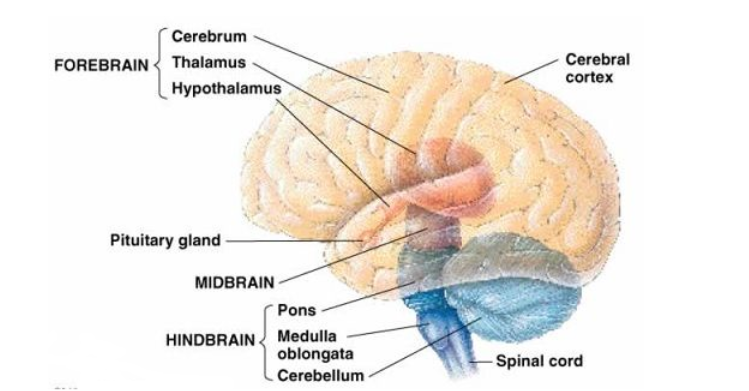
- The forebrain consists of cerebrum, thalamus and hypothalamus.
- Cerebrum forms the major part of the human brain.
- A deep cleft divides the cerebrum longitudinally into two halves, which are termed as the left and right cerebral hemispheres.
- The hemispheres are connected by a tract of nerve fibres called corpus callosum.
- The layer of cells which covers the cerebral hemisphere is called cerebral cortex and is thrown into prominent folds.
- The cerebral cortex is referred to as the grey matter due to its greyish appearance.The neuron cell bodies are concentrated here giving the colour.
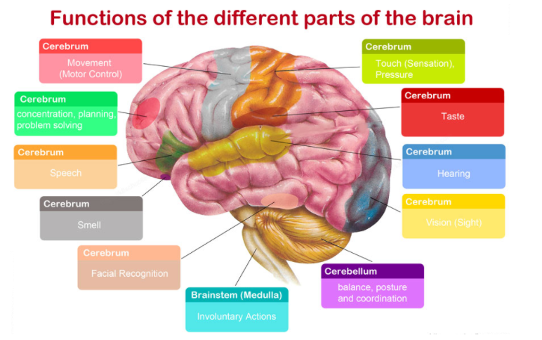
- The cerebral cortex contains motor areas, sensory areas and large regions that are neither clearly sensory or motor in function.
- These regions called association areas are responsible for complex functions like intersensory associations, memory and communication.
- Fibres of the tracts are covered with the myelin sheath, which constitute the inner part of the cerebral hemisphere.
- They give an opaque white appearance to the layer and, hence, are called the white matter.
- The cerebrum wraps around a structure called thalamus, which is a major coordinating centre for sensory and motor signalling.
- Another very important part of the brain called hypothalamus lies at the base of the thalamus.
- The hypothalamus contains a number of centres which control body temperature, urge for eating and drinking.
- It also contains several groups of neurosecretory cells, which secrete hormones called hypothalamic hormones.
- The inner parts of cerebral hemispheres and a group of associated deep structures like amygdala, hippocampus, etc., form a complex structure called the limbic lobe or limbic system.
- Along with the hypothalamus, it is involved in the regulation of sexual behaviour, expression of emotional reactions (e.g., excitement, pleasure, rage and fear), and motivation.
MIDBRAIN (Mesencephalon)
- The midbrain is located between the thalamus/hypothalamus of the forebrain and pons of the hindbrain. A canal called the cerebral aqueduct passess through the midbrain. The dorsal portion of the midbrain consists mainly of four round swellings (lobes) called corpora quadrigemina.
- Mid brain receives and integrates auditory, visual and tactile input.
HINDBRAIN
- The hindbrain comprises pons, cerebellum and medulla (also called the medulla oblongata).
- Pons consists of fibre tracts that interconnect different regions of the brain.
- Cerebellum has a very convoluted surface in order to provide the additional space for many more neurons.
- The medulla of the brain is connected to the spinal cord.
- The medulla contains centres which control respiration, cardiovascular reflexes and gastric secretions.
- Three major regions make up the brainstem; midbrain, pons and medulla oblongata. Brain stem forms the connections between the brain and spinal cord.
MENINGES
- They are protective membranous coverings of the brain and spinal cord. Meninges are three in number. They are dura mater, arachnoid and pia mater.
- Dura Mater- It is an outer thick layer of fibrous tissue that lines the cranial cavity. Dura mater resists the movement of the brain inside the cranium so that stretching and breaking of blood vessels do not occur.
- Arachnoid - It is a middle meninx, which is thin, webby, porous.
- Subdural space between the dura mater and arachnoid is filled with fluid which is not cerebrospinal fluid.
- Pia Mater - It is moderately tough inner meninx which is highly vascularised and pigmented.
- The space between the arachnoid and pia mater is called subarachnoid space. It contains cerebrospinal spinal fluid and connective tissue strands.

REFLEX ACTION AND REFLEX ARC
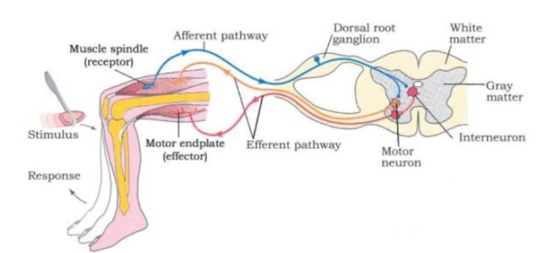
- You must have experienced a sudden withdrawal of a body part which comes in contact with objects that are extremely hot, cold pointed or animals that are scary or poisonous. The entire process of response to a peripheral nervous stimulation that occurs involuntarily, i.e., without conscious effort or thought and requires the involvement of a part of the central nervous system is called a reflex action.
- The reflex pathway comprises at least one afferent neuron (receptor) and one efferent (effector or excitor) neuron appropriately arranged in a series.
- The afferent neuron receives signal from a sensory organ and transmits the impulse via a dorsal nerve root into the CNS (at the level of spinal cord).
- The efferent neuron then carries signals from CNS to the effector. The stimulus and response thus forms a reflex arc.
-
COMPONENTS OF BRAIN

PERIPHERAL NERVOUS SYSTEM
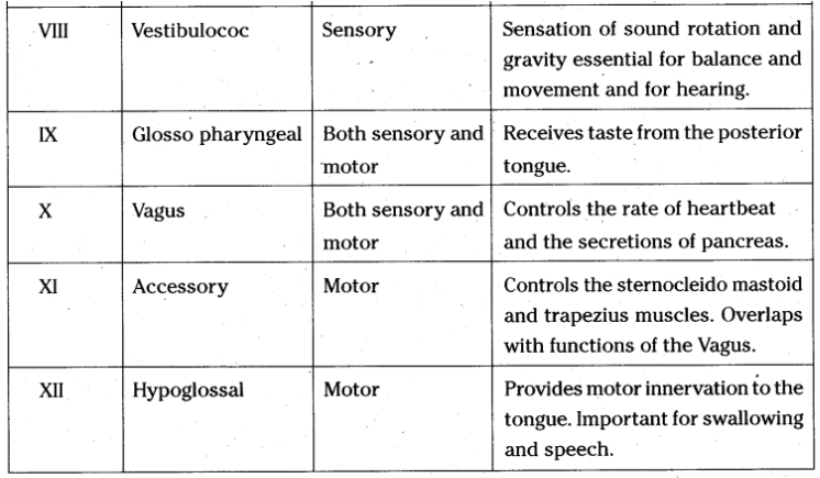
- It is formed of nerves extending between CNS and body tissues.
- On the basis of their origin, nerves are of two types:
I.Cranial nerves which arise from the brain.
II.Spinal nerves which arise from spinal cord. - Spinal nerves
Spinal nerves arise from gray matter of the spinal cord. There are 31 pairs of spinal nerves in man. All spinal nerves are mixed. The spinal nerves in man are divided into groups.- Cervical(C) → 8 pairs.
- Thoracic(T) → 12 pairs.
- Lumbar(L) → 5 pairs.
- Sacral(S) → 5 pairs.
- Coccygeal (CO) → 1 pair.
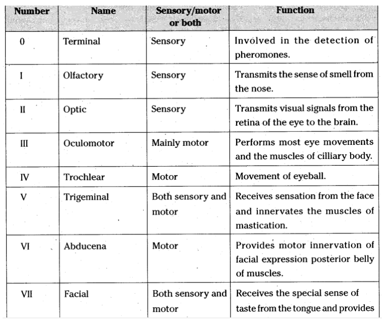
- Cranial nerves
- 10 pairs of cranial nerves are present in anamniotes(fishes and amphibians).
- 12 pairs of cranial nerves are present in an amniotes (reptiles, birds and mammals).
STRUCTURE OF BRAIN

Fore Brain
- The forebrain consists of cerebrum, thalamus and hypothalamus.
- Cerebrum forms the major part of the human brain.
- A deep cleft divides the cerebrum longitudinally into two halves, which are termed as the left and right cerebral hemispheres.
- The hemispheres are connected by a tract of nerve fibres called corpus callosum.
- The layer of cells which covers the cerebral hemisphere is called cerebral cortex and is thrown into prominent folds.
- The cerebral cortex is referred to as the grey matter due to its greyish appearance.The neuron cell bodies are concentrated here giving the colour.
- The cerebral cortex contains motor areas, sensory areas and large regions that are neither clearly sensory or motor in function.
- These regions called association areas are responsible for complex functions like intersensory associations, memory and communication.
- Fibres of the tracts are covered with the myelin sheath, which constitute the inner part of the cerebral hemisphere.
- They give an opaque white appearance to the layer and, hence, are called the white matter.
- The cerebrum wraps around a structure called thalamus, which is a major coordinating centre for sensory and motor signalling.
- Another very important part of the brain called hypothalamus lies at the base of the thalamus.
- The hypothalamus contains a number of centres which control body temperature, urge for eating and drinking.
- It also contains several groups of neurosecretory cells, which secrete hormones called hypothalamic hormones.
- The inner parts of cerebral hemispheres and a group of associated deep structures like amygdala, hippocampus, etc., form a complex structure called the limbic lobe or limbic system.
- Along with the hypothalamus, it is involved in the regulation of sexual behaviour, expression of emotional reactions (e.g., excitement, pleasure, rage and fear), and motivation.
MIDBRAIN (Mesencephalon)
- The midbrain is located between the thalamus/hypothalamus of the forebrain and pons of the hindbrain. A canal called the cerebral aqueduct passess through the midbrain. The dorsal portion of the midbrain consists mainly of four round swellings (lobes) called corpora quadrigemina.
- Mid brain receives and integrates auditory, visual and tactile input.
HINDBRAIN
- The hindbrain comprises pons, cerebellum and medulla (also called the medulla oblongata).
- Pons consists of fibre tracts that interconnect different regions of the brain.
- Cerebellum has a very convoluted surface in order to provide the additional space for many more neurons.
- The medulla of the brain is connected to the spinal cord.
- The medulla contains centres which control respiration, cardiovascular reflexes and gastric secretions.
- Three major regions make up the brainstem; midbrain, pons and medulla oblongata. Brain stem forms the connections between the brain and spinal cord.

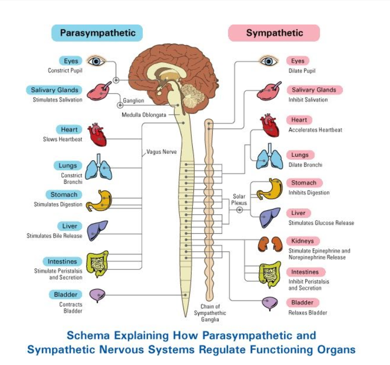
AUTONOMIC NERVOUS SYSTEM/VISCERAL NERVOUS SYSTEM
- It is the part of the peripheral nervous system that comprises the whole complex of nerves, fibers, ganglia and plexuses by which impulses travel from the central nervous system to the viscera and from the viscera to the CNS.
- ANS consists of two divisions: sympathetic and parasympathetic.
REFLEX ACTION AND REFLEX ARC
- You must have experienced a sudden withdrawal of a body part which comes in contact with objects that are extremely hot, cold pointed or animals that are scary or poisonous. The entire process of response to a peripheral nervous stimulation that occurs involuntarily, i.e., without conscious effort or thought and requires the involvement of a part of the central nervous system is called a reflex action.
- The reflex pathway comprises at least one afferent neuron (receptor) and one efferent (effector or excitor) neuron appropriately arranged in a series.
- The afferent neuron receives signal from a sensory organ and transmits the impulse via a dorsal nerve root into the CNS (at the level of spinal cord).
- The efferent neuron then carries signals from CNS to the effector. The stimulus and response thus forms a reflex arc.
DISEASES OF NERVOUS SYSTEM
PERIPHERAL NERVOUS SYSTEM
- It is formed of nerves extending between CNS and body tissues.
- On the basis of their origin, nerves are of two types:
I.Cranial nerves which arise from the brain.
II.Spinal nerves which arise from spinal cord. - Spinal nerves
Spinal nerves arise from gray matter of the spinal cord. There are 31 pairs of spinal nerves in man. All spinal nerves are mixed. The spinal nerves in man are divided into groups.- Cervical(C) → 8 pairs.
- Thoracic(T) → 12 pairs.
- Lumbar(L) → 5 pairs.
- Sacral(S) → 5 pairs.
- Coccygeal (CO) → 1 pair.
- Cranial nerves
- 10 pairs of cranial nerves are present in anamniotes(fishes and amphibians).
- 12 pairs of cranial nerves are present in an amniotes (reptiles, birds and mammals).


SENSORY RECEPTION AND PROCESSING
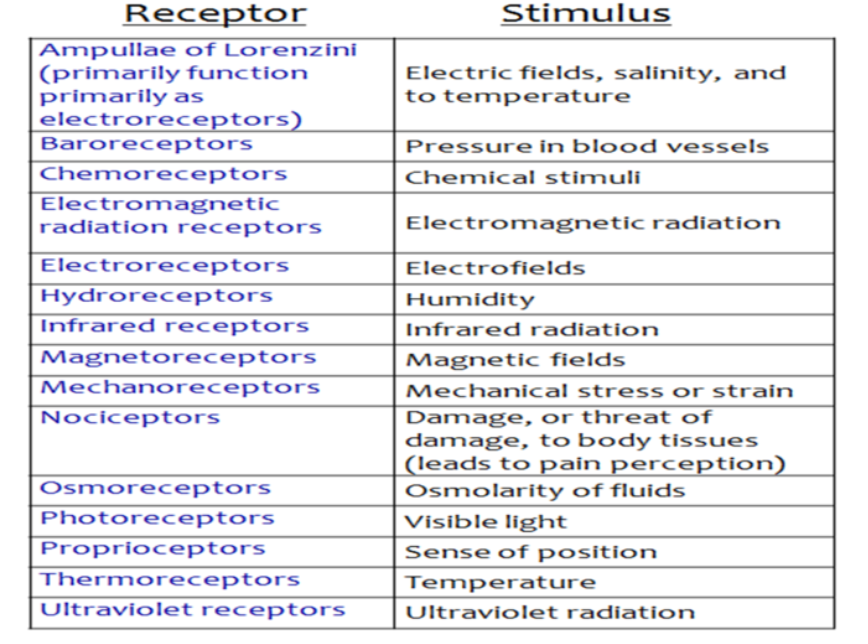
- The sensory organs detect all types of changes in the environment and send appropriate signals to the CNS, where all the inputs are processed and analysed. Signals are then sent to different parts/ centres of the brain. This is how you can sense changes in the environment.
- The nose contains mucus-coated receptors which are specialised for receiving the sense of smell and called olfactory receptors. These are made up of olfactory epithelium that consists of three kinds of cells. The neurons of the olfactory epithelium extend from the outside environment directly into a pair of broad bean-sized organs, called olfactory bulbs, which are extensions of the brain’s limbic system.
- Both nose and tongue detect dissolved chemicals. The chemical senses of gustation (taste) and olfactory (smell) are functionally similar and interrelated.
- The tongue detects tastes through taste buds, containing gustatory receptors. With each taste of food or sip of drink, the brain integrates the differential input from the taste buds and a complex flavour is perceived.
SENSE ORGANS
We smell things by our nose, taste by tongue, hear by ear and see objects by eyes.
AUTONOMIC NERVOUS SYSTEM/VISCERAL NERVOUS SYSTEM
- It is the part of the peripheral nervous system that comprises the whole complex of nerves, fibers, ganglia and plexuses by which impulses travel from the central nervous system to the viscera and from the viscera to the CNS.
- ANS consists of two divisions: sympathetic and parasympathetic.

EYE
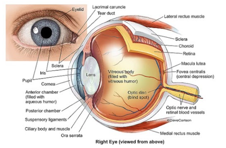
- Our paired eyes are located in sockets of the skull called orbits. A brief account of structure and functions of the human eye is given in the following sections.
- The adult human eyeball is nearly a spherical structure. The wall of the eyeball is composed of three layers.
- The external layer is composed of a dense connective tissue and is called the sclera.
- The anterior portion of this layer is called the cornea.
- The middle layer, choroid, contains many blood vessels and looks bluish in colour.
- The choroid layer is thin over the posterior two-thirds of the eyeball, but it becomes thick in the anterior part to form the ciliary body.
- The ciliary body itself continues forward to form a pigmented and opaque structure called the iris which is the visible coloured portion of the eye.
- The eyeball contains a transparent crystalline lens which is held in place by ligaments attached to the ciliary body.
- In front of the lens, the aperture surrounded by the iris is called the pupil. The diameter of the pupil is regulated by the muscle fibres of iris.
- The inner layer is the retina and it contains three layers of neural cells – from inside to outside – ganglion cells, bipolar cells and photoreceptor cells.
- There are two types of photoreceptor cells, namely, rods and cones.
- These cells contain the light-sensitive proteins called the photopigments.
- The daylight (photopic) vision and colour vision are functions of cones and the twilight (scotopic) vision is the function of the rods.
- The rods contain a purplish-red protein called the rhodopsin or visual purple, which contains a derivative of Vitamin A.
- In the human eye, there are three types of cones which possess their own characteristic photopigments that respond to red, green and blue lights.
- The sensations of different colours are produced by various combinations of these cones and their photopigments. When these cones are stimulated equally, a sensation of white light is produced.
- The optic nerves leave the eye and the retinal blood vessels enter it at a point medial to and slightly above the posterior pole of the eyeball.
- Photoreceptor cells are not present in that region and hence it is called the blind spot.
- At the posterior pole of the eye lateral to the blind spot, there is a yellowish pigmented spot called macula lutea with a central pit called the fovea.
- The fovea is a thinned-out portion of the retina where only the cones are densely packed. It is the point where the visual acuity (resolution) is the greatest.
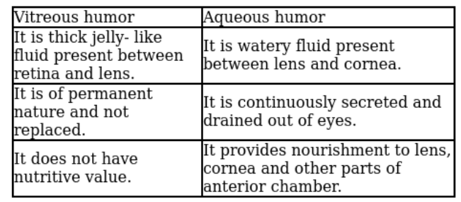
- The space between the cornea and the lens is called the aqueous chamber and contains a thin watery fluid called aqueous humor.
- The space between the lens and the retina is called the vitreous chamber and is filled with a transparent gel called vitreous humor.
- The light rays in visible wavelength focused on the retina through the cornea and lens generate potentials (impulses) in rods and cones.
- The photosensitive compounds (photopigments) in the human eye are composed of opsin (a protein) and retinal (an aldehyde of vitamin A).
- Light induces dissociation of the retinal from opsin resulting in changes in the structure of the opsin.This causes membrane permeability changes.
- As a result, potential differences are generated in the photoreceptor cells. This produces a signal that generates action potentials in the ganglion cells through the bipolar cells.
- These action potentials (impulses) are transmitted by the optic nerves to the visual cortex area of the brain, where the neural impulses are analysed and the image formed on the retina is recognised based on earlier memory and experience.
PARTS OF EYE
MECHANISM OF VISION
DISEASES OF NERVOUS SYSTEM
EAR
- The ears perform two sensory functions, hearing and maintenance of body balance.
- Anatomically, the ear can be divided into three major sections called the outer ear, the middle ear and the inner ear.
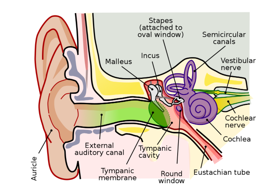
- The outer ear consists of the pinna and external auditory meatus (canal).
- The external auditory meatus leads inwards and extends up to the tympanic membrane (the eardrum).
- There are very fine hairs and wax-secreting glands in the skin of the pinna and the meatus.
- The tympanic membrane is composed of connective tissues covered with skin outside and with mucus membrane inside.
- The middle ear contains three ossicles called malleus, incus and stapes which are attached to one another in a chain-like fashion.
- The malleus is attached to the tympanic membrane and the stapes is attached to the oval window of the cochlea.
- The ear ossicles increase the efficiency of transmission of sound waves to the inner ear.
- An Eustachian tube connects the middle ear cavity with the pharynx. The Eustachian tube helps in equalising the pressures on either side of the eardrum.
- The fluid-filled inner ear called labyrinth consists of two parts, the bony and the membranous labyrinths.
- The bony labyrinth is a series of channels.
- Inside these channels lies the membranous labyrinth, which is surrounded by a fluid called perilymph.
- The membranous labyrinth is filled with a fluid called endolymph.The coiled portion of the labyrinth is called cochlea.
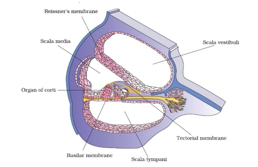
- The membranes constituting cochlea, the reissner’s and basilar, divide the surrounding perilymph filled bony labyrinth into an upper scala vestibuli and a lower scala tympani.
- The space within cochlea called scala media is filled with endolymph.
- At the base of the cochlea, the scala vestibuli ends at the oval window, while the scala tympani terminates at the round window which opens to the middle ear.
- The organ of corti is a structure located on the basilar membrane which contains hair cells that act as auditory receptors.
- The hair cells are present in rows on the internal side of the organ of corti.
- The basal end of the hair cell is in close contact with the afferent nerve fibres.
- A large number of processes called stereocilia are projected from the apical part of each hair cell.
- Above the rows of the hair cells is a thin elastic membrane called tectorial membrane.
- The inner ear also contains a complex system called vestibular apparatus, located above the cochlea.
- The vestibular apparatus is composed of three semicircular canals and the otolith (macula is the sensory part of saccule and utricle).
- Each semicircular canal lies in a different plane at right angles to each other.
- The membranous canals are suspended in the perilymph of the bony canals.
- The base of canals is swollen and is called ampulla, which contains a projecting ridge called crista ampullaris which has hair cells.
- The saccule and utricle contain a projecting ridge called macula.
- The crista and macula are the specific receptors of the vestibular apparatus responsible for maintenance of balance of the body and posture.
- The external ear receives sound waves and directs them to the eardrum.
- The eardrum vibrates in response to the sound waves and these vibrations are transmitted through the ear ossicles (malleus, incus and stapes) to the oval window.
- The vibrations are passed through the oval window onto the fluid of the cochlea, where they generate waves in the lymph.
- The waves in the lymph induce a ripple in the basilar membrane.
- These movements of the basilar membrane bend the hair cells, pressing them against the tectorial membrane.
- As a result, nerve impulses are generated in the associated afferent neurons.
- These impulses are transmitted by the afferent fibres via auditory nerves to the auditory cortex of the brain, where the impulses are analysed and the sound is recognised.
MECHANISM OF HEARING
SENSORY RECEPTION AND PROCESSING
SENSE ORGANS
We smell things by our nose, taste by tongue, hear by ear and see objects by eyes.
- The nose contains mucus-coated receptors which are specialised for receiving the sense of smell and called olfactory receptors. These are made up of olfactory epithelium that consists of three kinds of cells. The neurons of the olfactory epithelium extend from the outside environment directly into a pair of broad bean-sized organs, called olfactory bulbs, which are extensions of the brain’s limbic system.
- Both nose and tongue detect dissolved chemicals. The chemical senses of gustation (taste) and olfactory (smell) are functionally similar and interrelated.
- The tongue detects tastes through taste buds, containing gustatory receptors. With each taste of food or sip of drink, the brain integrates the differential input from the taste buds and a complex flavour is perceived.

SUMMARY
- The neural system coordinates and integrates functions as well as metabolic and homeostatic activities of all the organs.
- Neurons, the functional units of the neural system, are excitable cells due to a differential concentration gradient of ions across the membrane.
- The electrical potential difference across the resting neuronal membrane is called the ‘resting potential’.
- The nerve impulse is conducted along the axon membrane in the form of a wave of depolarisation and repolarisation.
- A synapse is formed by the membranes of a presynaptic neuron and a postsynaptic neuron which may or may not be separated by a gap called synaptic cleft.
- Chemicals involved in the transmission of impulses at chemical synapses are called neurotransmitters.
- Human neural system consists of two parts : (i) the central neural system (CNS) and (ii) the peripheral neural system.
- The CNS consists of the brain and spinal cord.
- The brain can be divided into three major parts : (i) forebrain, (ii) midbrain and (iii) hindbrain.
- The forebrain consists of cerebrum, thalamus and hypothalamus.
- The cerebrum is longitudinally divided into two halves that are connected by the corpus callosum.
- A very important part of the forebrain called hypothalamus controls the body temperature, eating and drinking.
- Inner parts of cerebral hemispheres and a group of associated deep structures form a complex structure called limbic system which is concerned with olfaction, autonomic responses, regulation of sexual behaviour, expression of emotional reactions, and motivation.
- The midbrain receives and integrates visual, tactile and auditory inputs.
- The hindbrain comprises pons, cerebellum and medulla.
- The cerebellum integrates information received from the semicircular canals of the ear and the auditory system.
- The medulla contains centres, which control respiration, cardiovascular reflexes, and gastric secretions.
- Pons consist of fibre tracts that interconnect different regions of the brain.
- The entire process of involuntary response to a peripheral nervous stimulation is called reflex action.
- Information regarding changes in the environment is received by the CNS through the sensory organs which are processed and analysed. Signals are then sent for necessary adjustments.
- The wall of the human eyeball is composed of three layers.
- The external layer is composed of cornea and sclera. Inside the sclera is the middle layer, which is called the choroid.
- Retina, the innermost layer, contains two types of photoreceptor cells, namely rods and cones.
- The daylight (photopic) vision and colour vision are functions of cones and twilight (scotopic) vision is the function of the rods.
- The light enters through the cornea, the lens and the images of objects are formed on the retina.
- The ear can be divided into the outer ear, the middle ear and the inner ear. The middle ear contains three ossicles called malleus, incus and stapes.
- The fluid filled inner ear is called the labyrinth, and the coiled portion of the labyrinth is called cochlea.
- The organ of corti is a structure which contains hair cells that act as auditory receptors and is located on the basilar membrane.
- The vibrations produced in the eardrum are transmitted through the ear ossicles and oval window to the fluid-filled inner ear.
- Nerve impulses are generated and transmitted by the afferent fibres to the auditory cortex of the brain.
- The inner ear also contains a complex system located above the cochlea called vestibular apparatus.
- It is influenced by gravity and movements, and helps us in maintaining balance of the body and posture.
EYE
- Our paired eyes are located in sockets of the skull called orbits. A brief account of structure and functions of the human eye is given in the following sections.
- The adult human eyeball is nearly a spherical structure. The wall of the eyeball is composed of three layers.
- The external layer is composed of a dense connective tissue and is called the sclera.
- The anterior portion of this layer is called the cornea.
- The middle layer, choroid, contains many blood vessels and looks bluish in colour.
- The choroid layer is thin over the posterior two-thirds of the eyeball, but it becomes thick in the anterior part to form the ciliary body.
- The ciliary body itself continues forward to form a pigmented and opaque structure called the iris which is the visible coloured portion of the eye.
- The eyeball contains a transparent crystalline lens which is held in place by ligaments attached to the ciliary body.
- In front of the lens, the aperture surrounded by the iris is called the pupil. The diameter of the pupil is regulated by the muscle fibres of iris.
- The inner layer is the retina and it contains three layers of neural cells – from inside to outside – ganglion cells, bipolar cells and photoreceptor cells.
- There are two types of photoreceptor cells, namely, rods and cones.
- These cells contain the light-sensitive proteins called the photopigments.
- The daylight (photopic) vision and colour vision are functions of cones and the twilight (scotopic) vision is the function of the rods.
- The rods contain a purplish-red protein called the rhodopsin or visual purple, which contains a derivative of Vitamin A.
- In the human eye, there are three types of cones which possess their own characteristic photopigments that respond to red, green and blue lights.
- The sensations of different colours are produced by various combinations of these cones and their photopigments. When these cones are stimulated equally, a sensation of white light is produced.
- The optic nerves leave the eye and the retinal blood vessels enter it at a point medial to and slightly above the posterior pole of the eyeball.
- Photoreceptor cells are not present in that region and hence it is called the blind spot.
- At the posterior pole of the eye lateral to the blind spot, there is a yellowish pigmented spot called macula lutea with a central pit called the fovea.
- The fovea is a thinned-out portion of the retina where only the cones are densely packed. It is the point where the visual acuity (resolution) is the greatest.
- The space between the cornea and the lens is called the aqueous chamber and contains a thin watery fluid called aqueous humor.
- The space between the lens and the retina is called the vitreous chamber and is filled with a transparent gel called vitreous humor.
- The light rays in visible wavelength focused on the retina through the cornea and lens generate potentials (impulses) in rods and cones.
- The photosensitive compounds (photopigments) in the human eye are composed of opsin (a protein) and retinal (an aldehyde of vitamin A).
- Light induces dissociation of the retinal from opsin resulting in changes in the structure of the opsin.This causes membrane permeability changes.
- As a result, potential differences are generated in the photoreceptor cells. This produces a signal that generates action potentials in the ganglion cells through the bipolar cells.
- These action potentials (impulses) are transmitted by the optic nerves to the visual cortex area of the brain, where the neural impulses are analysed and the image formed on the retina is recognised based on earlier memory and experience.

PARTS OF EYE

MECHANISM OF VISION
Eye
- Eye ek ball like structure hai jo facial bones ki eye socket me located hai and it is richly supplied with nerves, blood vessels and muscles. Eyeball me 3 layers hoti hai :
- Fibrous tunic : yeah sabse outermost layer hai jo anteriorly cornea and posteriorly sclera se milkar banti hai. Cornea transparent coat hai jo colored iris ko cover karta hai. Cornea curved hai and issi curvature ke karan yeah incident light ko retina pe focus karne ka kaam bhi karta hai. Dusra kaam cornea ka hai - bahar ki air se directly oxygen receive karna. Cornea me blood supply nhi hoti. Aankhon me jo safed hissa dikhta hai vhi sclera kehlata hai jo ki mostly collagen fibers se bani hui layer hai. Sclera covers the entire eyeball except the cornea; it gives shape to the eyeball, makes it more rigid, protects its inner parts, and serves as a site of attachment for the extrinsic eye muscles.
- Vascular tunic : eyeball ki middle layer hai. It has 3 parts : choroid, ciliary body and iris.
- Posterior side par, sclera ke neeche choroid layer hoti hai jo ki highly vascularized hai. Iss layer me pigment cells yani ki melanocytes hote hai toh yeah deep brown color ki dikhti hai. Pigment ka role yeah hai ki aankho me aane wali stray light ko absorb karta hai taki aankho ke ander reflection na ho and light scatter na kare. Issi layer ka anterior portion ciliary body ban jata hai. Isme bhi pigment cells hote hai. Ciliary body ke 2 main parts hai ciliary processes and ciliary muscles.
- Ciliary process aqueous humor secrete karti hai and issi se suspensory ligaments attached hote hai jo in turn eye lens ko uski position me hold karke rakhte hai.
- Ciliary muscle ek circular smooth muscle hai jiske contraction and relaxation se eye lens ki shape change hoti hai and it helps in accommodation of the eye.
- Iris eye ka colored part hai jo ki cornea and lens ke beech located hai. It consists of melanocytes and circular and radial smooth muscle fibers. The amount of melanin in the iris determines the eye color. Iris ka main function hai eyeball me enter kar rhi light ki amount ko regulate karna and this is done through pupil. Pupil iris ke center me located hole hai jo ki dark blak dot ki tarah dikhta hai. Iris ki smooth circular and radial muscles hi light ke respect me iss pupil ke size ko control karte hai and determine karte hai ki kitni light eye me enter kar rhi hai.
- Posterior side par, sclera ke neeche choroid layer hoti hai jo ki highly vascularized hai. Iss layer me pigment cells yani ki melanocytes hote hai toh yeah deep brown color ki dikhti hai. Pigment ka role yeah hai ki aankho me aane wali stray light ko absorb karta hai taki aankho ke ander reflection na ho and light scatter na kare. Issi layer ka anterior portion ciliary body ban jata hai. Isme bhi pigment cells hote hai. Ciliary body ke 2 main parts hai ciliary processes and ciliary muscles.
- Retina eye ki innermost layer hai jo posterior 3 quarter me located hai. Retina me pigment layer and neural layers hoti hai. Pigment layer me toh vahi melanocytes hote hai and neural layer basically multilayered structure hai jo visual data ko process karta hai and then optic nerve ke through brain tak impulses bhejta hai.
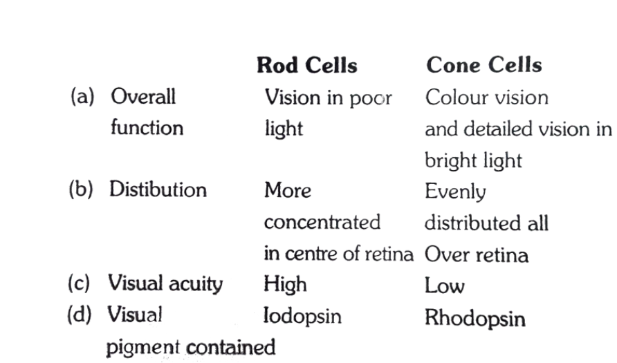
- Retina me hi 2 main type ke photoreceptors hote hai : rods (jo black and white vision ke liye responsible hote hai and dim light me activate honge) and cons (jo bright light me activate hote hai and colored vision ke liye responsible hote hai.).
- Retina me 120 million rods and 6 million cones hote hai. Three types of cones are present in the retina: (1) blue cones, which are sensitive to blue light, (2) green cones, which are sensitive to green light, and (3) red cones, which are sensitive to red light.
- Retina ka wo hissa jhan se optic nerve shuru hokr eyeball ko leave karti hai uss spot ko blind spot bola jata hai kyunki vhan pe na rod cells hote hai or na hi cone cells hote hai toh yhaan par image nahi bana sakti. Toh in case lens ne incident light ko iss spot pe focus kiya toh koi image nahi banegi.
- Eye ke retina ke bilkul posterior center me ek yellow spot hota hai jiske macula lutea bola jata hai. Issi me ek chota sa depression hota hai jiske fovea centralis kehte hai and iss fovea centralis me keval cones hi hote hai and yeah hissa highest visual acuity wala part hai yani ki iss part pe jo image banegi toh maximum clarity wali image banegi.
- Eye Lens eyeball ke ander ke hisse ko 2 cavities me divide karta hai : anterior cavity and vitreous chamber. Anterior cavity me aqueous humor naam ki fluid hoti hai jabki vitreous chamber me vitreous humor naam ki fluid hoti hai.
- Lighter medium se light jab denser medium me travel karti hai toh light rays bend hoti hai and issi ko hum refraction kehte hai. Right? Toh air se aane wali incident light jab eyeball me enter karti hai toh cornea usko refract karega and then eye lens (convex lens) firse refract karke usko retina pe focus karta hai. Retina pe form hone wali image inverted image banti hai.
- Accomodation of eye : Jab hum kisi najdeek ke object pe focus kar rahe hote hai toh lens or jyada curved ho jata hai jiske karna light rays ki jyada refraction hoti hai and it helps us to see nearby object. Dur ke object ko dekhte hue lens elongate hota hai and uska curvature comparatively less hota hai. Aankho ki yahi ability accommodation kehlati hai. Wo kam se kam duri jhan se aap clearly kisi najdeek ke object ko dekh sake wo near point of vision kehlata hai and for humans it is 10cm/4inch for the adult eye.
- Rods and cones me rhodopsin and cone pigments naam ke visual pigments hai, respectively. Yeah photopigments me 2 parts hai : opsin + retinal. Opsin ek glycoprotein hai jabki retinal, vit. A ka derivative hai. In sabhi visual pigments me retinal light absorbing part hai.
- Rods and cones ke pigments me, opsin part hi different hota hai. Even red, blue and green cones me bhi alag alag tarah ke opsin part hote hai. Yahi photopigments light ko respond karte hai and vision me inhi ka key role hai.
- Light ki absence kaho ya darkness me retinal apni cis form me hota hai. Yeah cis vahi chemistry ke cis and trans double bond wala cis hai. Isko hum cis=retinal kehte hai jab cis-retinal light ke ek photon ko absorb karta hai toh yeah cis se trans form me badal jata hai. Iss step ko hum isomerimerization kehte hai.
- Iss change ke baad photoreceptor me chemical changes (membrane ki ions ke liye permeability change hojati hai jaise neuron ke case me hui thi although mechanism is little bit different and wo syllabus me hai bhi nhi so wo won’t study that part into detail as of now.) hote hai jo action potential generate karte hai.
- Thodi hi der me trans-retinal, opsin part se completely detach hojata hai and iss step ko bleaching bola jata hai.
- Fir agli step me retinal isomerase naam ka enzyme, trans retinal ko firse cis retinal me badal deta hai and yeah cis retinal ab opsin ke sath firse bind karta hai. Iss fourth step ko regeneration bola jata hai and ab yeah functional photopigment light ke next photon ko absorb karne ke liye tyaar hojata hai.
Mechanism of Vision :
EAR
- The ears perform two sensory functions, hearing and maintenance of body balance.
- Anatomically, the ear can be divided into three major sections called the outer ear, the middle ear and the inner ear.
- The outer ear consists of the pinna and external auditory meatus (canal).
- The external auditory meatus leads inwards and extends up to the tympanic membrane (the eardrum).
- There are very fine hairs and wax-secreting glands in the skin of the pinna and the meatus.
- The tympanic membrane is composed of connective tissues covered with skin outside and with mucus membrane inside.
- The middle ear contains three ossicles called malleus, incus and stapes which are attached to one another in a chain-like fashion.
- The malleus is attached to the tympanic membrane and the stapes is attached to the oval window of the cochlea.
- The ear ossicles increase the efficiency of transmission of sound waves to the inner ear.
- An Eustachian tube connects the middle ear cavity with the pharynx. The Eustachian tube helps in equalising the pressures on either side of the eardrum.
- The fluid-filled inner ear called labyrinth consists of two parts, the bony and the membranous labyrinths.
- The bony labyrinth is a series of channels.
- Inside these channels lies the membranous labyrinth, which is surrounded by a fluid called perilymph.
- The membranous labyrinth is filled with a fluid called endolymph.The coiled portion of the labyrinth is called cochlea.
- The membranes constituting cochlea, the reissner’s and basilar, divide the surrounding perilymph filled bony labyrinth into an upper scala vestibuli and a lower scala tympani.
- The space within cochlea called scala media is filled with endolymph.
- At the base of the cochlea, the scala vestibuli ends at the oval window, while the scala tympani terminates at the round window which opens to the middle ear.
- The organ of corti is a structure located on the basilar membrane which contains hair cells that act as auditory receptors.
- The hair cells are present in rows on the internal side of the organ of corti.
- The basal end of the hair cell is in close contact with the afferent nerve fibres.
- A large number of processes called stereocilia are projected from the apical part of each hair cell.
- Above the rows of the hair cells is a thin elastic membrane called tectorial membrane.
- The inner ear also contains a complex system called vestibular apparatus, located above the cochlea.
- The vestibular apparatus is composed of three semicircular canals and the otolith (macula is the sensory part of saccule and utricle).
- Each semicircular canal lies in a different plane at right angles to each other.
- The membranous canals are suspended in the perilymph of the bony canals.
- The base of canals is swollen and is called ampulla, which contains a projecting ridge called crista ampullaris which has hair cells.
- The saccule and utricle contain a projecting ridge called macula.
- The crista and macula are the specific receptors of the vestibular apparatus responsible for maintenance of balance of the body and posture.
MECHANISM OF HEARING
- The external ear receives sound waves and directs them to the eardrum.
- The eardrum vibrates in response to the sound waves and these vibrations are transmitted through the ear ossicles (malleus, incus and stapes) to the oval window.
- The vibrations are passed through the oval window onto the fluid of the cochlea, where they generate waves in the lymph.
- The waves in the lymph induce a ripple in the basilar membrane.
- These movements of the basilar membrane bend the hair cells, pressing them against the tectorial membrane.
- As a result, nerve impulses are generated in the associated afferent neurons.
- These impulses are transmitted by the afferent fibres via auditory nerves to the auditory cortex of the brain, where the impulses are analysed and the sound is recognised.
Ear
- Ear 3 main regions me divisible hai : external ear, middle ear and inner ear.
- External ear me auricle, external auditory canal and eardrum aate hai.
- Pinna/auricle elastic cartilage ka structure hai jo surrounding se auditory waves ko collect karke, external auditory canal ke through unko eardrum tak direct karta hai.
- Tympanic membrane or eardrum is a thin, semitransparent partition between the external auditory canal and middle ear.
- External auditory canal me hairs and sweat glands hote hai. Hair, air se dust and large particulate matter ko filter karte hai.
- Sweat glands ko ceruminous glands bola jata hai and yeah earwax/cerumen secrete karte hai. The combination of hairs and cerumen helps prevent dust and foreign objects from entering the ear. Cerumen also prevents damage to the delicate skin of the external ear canal by water and insects.
- Middle ear me 3 ear bones hoti hai jinko auditory ossicles bhi bola jata hai. Yeah bones me bhi synovial joints hote hai.
- Hammer (malleus), anvil (incus) and stirrup (stapes) in bones ke naam hai.
- Malleus bone yani ki hammer shaped bone ka handle part, tympanic membrane se juda hua hota hai.
- Malleus se incus and incus se stapes bone judi hui hai. Stapes ka foot part oval window se attached hota hai.
- Oval window ke neeche hi ek round window bhi hoti hai. Middle ear ke anterior hisse me ek opening hoti hai jo eustachian tube me open hoti hai.
- Eustachian tube ko hi auditory tube bhi kehte hai. Yeah middle ear ko throat ke superior part se jodti hai. Iska function hai middle ear me air pressure ko atmospheric pressure ke equal rakhna.
- Internal ear ya inner ear ko labyrinth bhi kehte hai kyunki isme canals ki series hoti hai. Structurally dekhe toh 2 parts hai : bony labyrinth and membranous labyrinth.
- Bony labyrinth temporal bone ki cavity me situated hai and iske further 3 regions hai : semicircular canals, vestibule and cochlea.
- Bony labyrinth me perilymph naam ki fluid hoti hai and issi fluid me membranous labyrinth hai jo ki epithelial sacs and tubes ka structure hai and bony labyrinth wale hi components yani ki membranous semicircular canals, vestibule ans cochlea iska part hai. Isko aise yaad rakh skate ho ki membrane semicircular canal, vestibule and cochlea ke bahar same shape ka bony structure hai jiske bony labyrinth kehte hai. Membranous labyrinth me jo fluid hoti hai usko endolymph kaha jata hai.
- Vestibule bony labyrinth ka central oval shaped portion hai jisme 2 membranous sacs present hai jinko utricle and saccule kehte hai.
- Vestibule ke superior-posterior aspect se 3 semicircular canals nikalti hai. Har ek canal ka end swollen hota hai jiske ampulla bola jata hai.
- Vestibule ke anterior side cochlea hota hai jo ki ek bony spiral hai canal hai and iski shape snail ke shell se resemble karti hai. Issi ke ander cochlear duct hoti hai jo ki membranous portion hai.
- Cochlear duct ka hi dusra naam scala media hai and yeah endolymph se filled hai.
- Scala media ke uper wala chamber scala vestibuli kehlata hai jo oval window pe khatam hota hai. Scala media ke neeche wala channel scala tympani kehlata hai jo ki round window pe end hota hai.
- Scala vestibuli and scala tympani dono hi bony labyrinth ka part hai and yeah perilymph se filled hai.
- Scala vestibuli and scala tympani are completely separated by the cochlear duct, except for an opening at the apex of the cochlea, the helicotrema.
- Vestibular membrane, scala vestibuli nad cochlear duct ke beech me present hoti hai jabki basilar membrane cochlear duct and scala tympani ko separate karti hai.
- Basilar membrane pe spiral organs/organ of corti hota hai jo ki epithelial cells ki ek coiled sheet hai with a lot of hair cells. Yeah hair cells hi audio waves ko receive karne ka kaam karte hai. Har ek hair cell ki tip pe stereocilia hote hai jo cochlear duct ki endolymph me extend karte hai.
- Despite their name, stereocilia are actually long, hairlike microvilli arranged in several rows of graded height.
- Tectorial membrane ek flexible gelatinous membrane hai jo spiral organs ke hair cells ko cover karta hai and stereocilia ke ends tectorial membrane me hi embedded hote hai jabki hair cells ki basal body basilar membrane pe situated hai.
- Inner hair cells hi sound ke mechanical vibrations ko electrical signals/nerve impulses me convert karte hai and in nerve impulses ko nerves brain ke auditory center me lekr jati hai jhan inki processing hoti hai.
- Auricle/pinna sound waves ko collect karke external auditory canal me bhejta hai.
- Fir sound waves tympanic membrane se takrati hai and usko vibrate karwati hai.
- Eardrum se ear ossicles connected hai toh jab eardrum vibrate karta hai toh ear ossicles bhi vibrate karte hai.
- 3rd ear ossicle yani ki stapes, oval window ki membrane se connected hai toh jab stapes vibrate karti hai toh oval window vibrate karti hai. Ear ossicles basically sound waves ko amplify karte hai.
- Yaad karo ki oval window ke ander perilymph hai toh jab oval window vibrate karti hai toh scala vestibuli ki perilymph fluid move karti hai.
- Scala vestibuli se pressure waves scala tympani tak jati hai and we know ki scala tympani round window me end hoti hai. Toh finally ab round window vibrate karti hai.
- Scala vestibuli and scala tympani me sound waves jab perilymph me vibrations paida karti hai toh eventually yeah vestibular membrane ko vibrate karti hai jiske karan cochlear duct me endolymph move karti hai.
- Iske karan ab basilar membrane me vibrations hote hai jo spiral organs ko tectorial membrane ke against move karte hai. Iske karan stereocilia bend hojate hai and this leads to generation of nerve impulses in cochlear nerve fibers. Fir yeah impulses brain tak nerves ke through jati hai and brain me respective areas iss sensory information ko decode karte hai.
- Otolith ka matlab hai oval shaped calcareous bodies, simply bole toh chote patthar ya granules.
- Utricle and saccule otolith organs hai yani ki inme yeah granules present hai. Inn organs ka thickened hissa macula kehlata hai jinme ki linear acceleration/deceleration and head ki position ko detect karne ke liye receptors hote hai.
- Yeah sensory receptors bhi basically hair cells hi hote hai jinke surface pe bahut sare stereocilia + 1 kinocilium (bada stereocilia) hota hai.
- Utricle and saccule ke maculae ek dusre ke perpendicular hote hai jiske karan inka functional role bhi alag alag hote hai.
- Utricle ka macula horizontal hota hai jabki saccule ka macula vertically oriented hota hai. Iska functional significance yeah hai ki utricle linear acceleration/deceleration that occurs in horizontal direction and change in position of head (forward and backward tilting of head) ko sense karta hai jabki saccule verticle direction me linear acceleration me change ko sense karta hai jaise ki elevator me upar neeche aatey jatey hue.
- Head ko tilt karte hue otoliths ki position change hoti hai jo stereocilia and kinocilium ko bend karte hai and issi bending se actional potential generate hota hai jo nerves ke through corresponding brain areas tak bhej di jati hai.
- 3 semicircular ducts ek dusre ke perpendicular lie karti hai and yeah rotational acceleration/deceleration me changes ko sense karti hai.
- 3 canals ka end part swollen hota hai jisko ampulla bola jata hai.
- Ampulla me ek chota sa ubhaar ya elevation hota hai jisko crista bolte hai. Inhi crista pe hair cells located hote hai along with supporting cells. In hair cells me bhi stereocilia+kinocilium hota hai.
- Cupula naam ki gelatinous covering, in cristae ko cover karti hai. Jab rotate kiya hata hai toh semicircular canals jo ki attached hai wo bhi move karti hai lekin in semicircular me jo endolymph hoti hai wo inertia of rest ke karan move ni kar pati and yeah hair cells ko ben karwati hai.
- Head ki rotation ki opposite direction me cilia bend karte hai and action potential generate karte hai and yeah nerves ke through again brain me jakr decode hote hai.
Physiology of Hearing :
Equilibrium
Otolith Organs : Utricle & SacculeSemicircular Ducts
SUMMARY
- The neural system coordinates and integrates functions as well as metabolic and homeostatic activities of all the organs.
- Neurons, the functional units of the neural system, are excitable cells due to a differential concentration gradient of ions across the membrane.
- The electrical potential difference across the resting neuronal membrane is called the ‘resting potential’.
- The nerve impulse is conducted along the axon membrane in the form of a wave of depolarisation and repolarisation.
- A synapse is formed by the membranes of a presynaptic neuron and a postsynaptic neuron which may or may not be separated by a gap called synaptic cleft.
- Chemicals involved in the transmission of impulses at chemical synapses are called neurotransmitters.
- Human neural system consists of two parts : (i) the central neural system (CNS) and (ii) the peripheral neural system.
- The CNS consists of the brain and spinal cord.
- The brain can be divided into three major parts : (i) forebrain, (ii) midbrain and (iii) hindbrain.
- The forebrain consists of cerebrum, thalamus and hypothalamus.
- The cerebrum is longitudinally divided into two halves that are connected by the corpus callosum.
- A very important part of the forebrain called hypothalamus controls the body temperature, eating and drinking.
- Inner parts of cerebral hemispheres and a group of associated deep structures form a complex structure called limbic system which is concerned with olfaction, autonomic responses, regulation of sexual behaviour, expression of emotional reactions, and motivation.
- The midbrain receives and integrates visual, tactile and auditory inputs.
- The hindbrain comprises pons, cerebellum and medulla.
- The cerebellum integrates information received from the semicircular canals of the ear and the auditory system.
- The medulla contains centres, which control respiration, cardiovascular reflexes, and gastric secretions.
- Pons consist of fibre tracts that interconnect different regions of the brain.
- The entire process of involuntary response to a peripheral nervous stimulation is called reflex action.
- Information regarding changes in the environment is received by the CNS through the sensory organs which are processed and analysed. Signals are then sent for necessary adjustments.
- The wall of the human eyeball is composed of three layers.
- The external layer is composed of cornea and sclera. Inside the sclera is the middle layer, which is called the choroid.
- Retina, the innermost layer, contains two types of photoreceptor cells, namely rods and cones.
- The daylight (photopic) vision and colour vision are functions of cones and twilight (scotopic) vision is the function of the rods.
- The light enters through the cornea, the lens and the images of objects are formed on the retina.
- The ear can be divided into the outer ear, the middle ear and the inner ear. The middle ear contains three ossicles called malleus, incus and stapes.
- The fluid filled inner ear is called the labyrinth, and the coiled portion of the labyrinth is called cochlea.
- The organ of corti is a structure which contains hair cells that act as auditory receptors and is located on the basilar membrane.
- The vibrations produced in the eardrum are transmitted through the ear ossicles and oval window to the fluid-filled inner ear.
- Nerve impulses are generated and transmitted by the afferent fibres to the auditory cortex of the brain.
- The inner ear also contains a complex system located above the cochlea called vestibular apparatus.
- It is influenced by gravity and movements, and helps us in maintaining balance of the body and posture.
