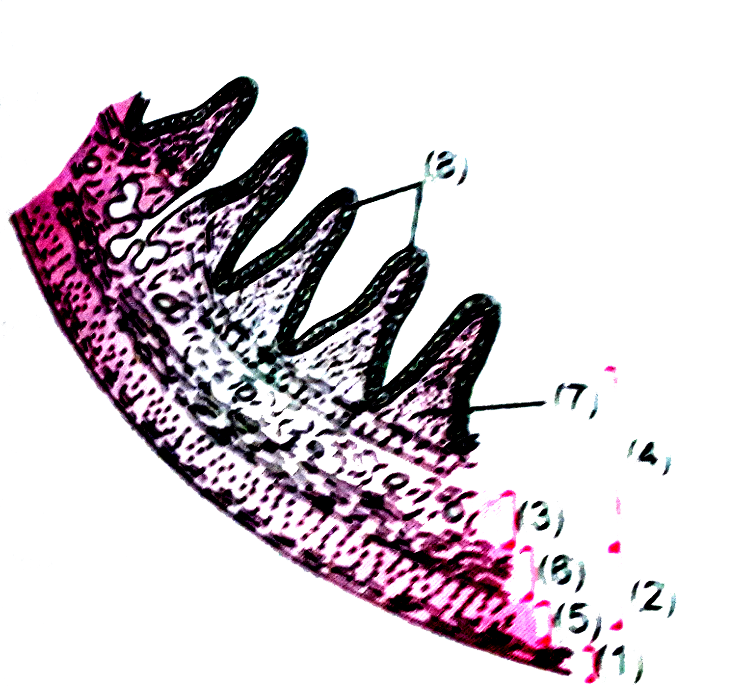
Figure depicts microsopic structure of the intestinal wall of alimentary canal. Study the figure and answer the following question?
(i) The wall of alimentary canal consists of 4 main concentric coats labelled as 1,2,3 and 4. Name these coats.
(ii) The coat marked 2 has two layers marked as 5 and 6. Between these two layers is a network of nerve cells and para-sympathetic nerve fibres called..... It controls the .......
(iii) The coat marked 3 is highly vasucular connective tissue having another network of nerve cells and sympathetic nerve fibres called...... . it controls the secretion of ........
(iv) Name the parts labelled as 7 and 8

Video Solution
Text Solution
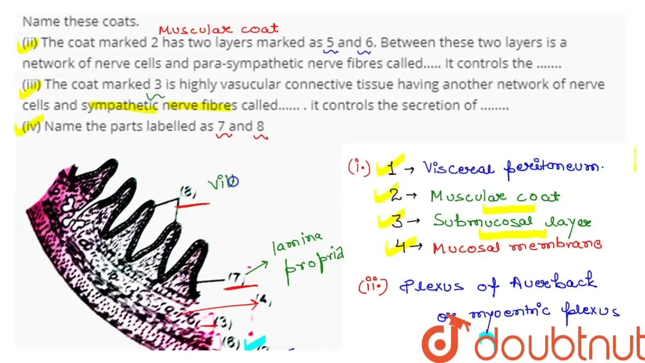
(i) 1. vescral peritoneum or serosa. 2. Muscular coat. 3. Sub-mucosa 4. Mucosa or mucous membrane.
(ii) Plexus of aueback of myoentric plexus, muscular contractions.
(iii) Plexus of Meissner or submucosal plexus, intestinal juice.
(iv) 7. Lamina propria 8. villi
|
Updated on:21/07/2023
Related Playlists
- BOOK - PRADEEPCHAPTER - DIGESTION AND ABSORPTIONEXERCISE - ADDITIONAL QUESTIONS ( LONG ANSWER QUESTIONS )26videos
DIGESTION AND ABSORPTION
- BOOK - PRADEEPCHAPTER - DIGESTION AND ABSORPTIONEXERCISE - ANALYTICAL QUESTIONS20videos
DIGESTION AND ABSORPTION
- BOOK - PRADEEPCHAPTER - DIGESTION AND ABSORPTIONEXERCISE - ADDITIONAL QUESTIONS (VERY SHORT ANSWER QUESTIONS )54videos
DIGESTION AND ABSORPTION
- BOOK - PRADEEPCHAPTER - CHEMICAL COORDINATION AND INTEGRATIONEXERCISE - Model Test Paper26videos
CHEMICAL COORDINATION AND INTEGRATION
- BOOK - PRADEEPCHAPTER - ECOSYSTEMEXERCISE - Assertion and reasoning16videos
ECOSYSTEM
Similar Questions
Alimentary Canal
View SolutionOutermost layer in the wall of alimentary canal is
View SolutionMeissner’s plexus is a network of nerve cells and sympathetic nerve fibres which control secretion of
View SolutionLamina propria in the alimentary canal consists of
View SolutionMucosa membrane of alimentary canal consists of
View SolutionAlimentary canal wall contains
View Solutionwhich of the following takes foods but has no alimentary canal ?
View SolutionThe alimentary canal in human has length of
View SolutionThe outermost layer of the wall of alimentary canal is made up of
View SolutionThe alimentary canal in human has length of
View Solution


