Describe various types of epithelial tissues with the help of labelled diagrams.
Epithelial tissues. They make inner or outer or both linings of different organs of body. They may be ectodermal, endodermal or mesodermal in origin. The term epithelium was given by Ruysch.
Types of epithelial tissues :
A. Simple epithelial tissue.
B. Complex or compound epithelial tissue.
A. Simple epithelial tissues. They have one layer of cells. The simple epithelial tissues are of the following five types :
1. Squamous epithelium.
2. Cuboidal epithelium.
3. Columnar epithelium.
4. Ciliated epithelium.
5. Pseudo stratified epithelium.
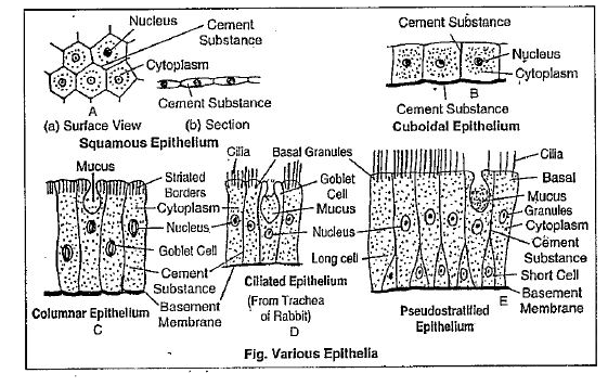
1. Squamous epithelium. Location. It makes lining of Bowman.s capsule, alveoli oflungs, skin castings of frog, membranous labyrinth ofinternal ear. The squamous epithelium which makes fining of blood vessels is called endothelium and which makes lining to body cavity is called mesothelium. Endothelium and mesothelium have wavy margin and called Tesseiated epithelium.
Characters.
(a) Cells are polygonal and isodiametric.
(b) Cells are closely packed like the tiles of a floor and that is why it is also called Pavement membrane.
(c) They have no vacuoles and intercellular spaces.
(d) Nucleus is flat and conspicuous.
(e) They have dense cytoplasm and rest on basement membrane.
(f) The .cells appear to be rectangular in V.S.
2. Cuboidal epithelium. Location. It makes lining of
(a) Ovaries as germinal epithelium.
(b) Seminiferous tubules as germinal epithelium.
(c) Limbs ofuriniferous tubules. Characters.
(i) Cells are cuboidal and closely packed.
(ii) The cells have rounded nuclei which are present in the centre.
(iii) Cytoplasm is dense and has no vacuoles.
(iv). It rests on basement membrane.
(v) Certain cells have evaginations called microvilli to increase the absorptive surface area. Due to these microvilli the cells have brush.border.
3. Columnar epithelium.
Location. It is present in enteric epithelium of stomach, intestine, gall bladder, gastric glands etc.
Characters:
(a) The cells are columnar or pillar like and generally closely packed.
(b) The cells have vacuolated cytoplasm.
(c) ThenuC!eus is oval and not present in centre.
(d) Certain cells have mucus vesicles, such cells are called goblet cells.
(e) Certain cells ofenteric epithelium ofifeum have microvilli which increase the surface for absorption of food.
(f) The columnar epithelium rests on basement membrane.
4. Ciliated epithelium. These cells have cilia on free surface. It is of two types :
(a) Cuboidal ciliated epithelium. It makes lining ofuriniferous tubules. The cells have cuboidal appearance. They have cilia on exposed surface which is towards lumen of uriniferous tubules. These cilia arise from basal granule.
(b) Columnar ciliated epithelium. It makes lining oflungs, oviducts, bronchi, bronchioles.
Characters.
(i) Cells are columnar.
(ii) Cells have cilia at free surface.
(iii) The ciliated epithelial cells are interrupted by mucus gland cells.
 (iv) The cells have oval nuclei which are eccentric.
(v) Cytoplasm is vacuolated.
(vi) Replacement cells are present.
5. Pseudostratifieil eptilzelium :
Types
(a) Pseudostratijied columnar epithelium. It appears to be two layered but actually it is one layered due to the presence of nuclei at different levels in large and small cells.
Location It makes lining of urethra, parotid salivary glands etc.
Characters.
(i) The larger cells are columnar and smaller cells triangular:
(ii) The larger cells.have· oval nuclei.
(iii) The smaller cells have round nuclei.
(iv) The larger columnar cells may be goblet cells.
(b) Pseudostratijied ciliated epitlzelium.
Location. It makes lining of trachea, large bronchi etc. Characters.
(i) The larger cells have cilia on free surface which arise from basal granules.
(ii) The cqlumnar cells have oval nuclei.
(iii) The small triangular cells have rounded nuclie.
(. ) The goblet cells are also present.
B. Compound pithelium. It has many layers. Compound epithelium is of two types . viz. staratified compound epithelium , and transitional compound epithelium.
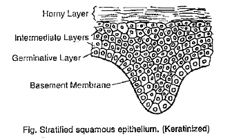
(a) Keratinized stratified squamous epithelium. It is the epidermis of mammals. Its characters are :
l. The lowermost layer is wavy and has columnar cells. These cells have the ability of dividing. It is called malpighian layer. The cells of this layer divide and shift outwardly.

2. The layers outer to stratum germinativum have flat cells.
3. The outermost few layers become dead due to the deposition of keratin and the process is called keratinization.
4. All layers between stratum malpighi and outer dead cells are called transitional layers.
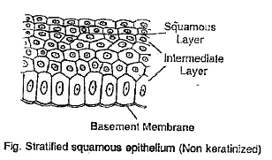
(b)Non-Keratinized squamous epithelium
Location. It makes lining of roof of buccal cavity, pharynx, oesophagus.
Characters. It is like keratinized stratified squamous epithelium but outer layers have flat living cells. There is no deposition of keratin in the cytoplasm.

(b) Stratified cuboidal epithelium. It makes lining of salivruy pancreatic ducts. It is also present in epidermis of scoliodon.
Characters.
1. The innermost layer is stratum malpighi.
2. It has columnar or low columnar cells.
3. The. outermost layer has cuboidal cells.
4. The transitional layers have flat or polygonal cells.
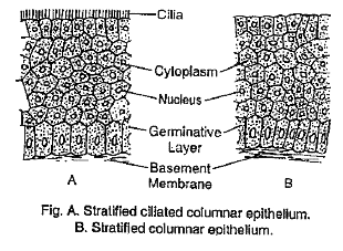
(c) Stratified columnar epithelium.
Location. It makes lining of epiglottis and some parts of urethra.
Characters.
(1) The stratliin germinativum has columnar cells which rest on the-basement membrane.

(ii) The outermost layer has also columnar cells.
(iii) Cells are living.
(iv) Transitional layers have polygonal cells.
(d) Stratified ciliated columnar epithelium. It makes lining oflarynx and upper part of soft palate.
Characters. 1. Stratum malpighi has columnar cells. 2. The outermost layer has columnar ciliated cells. 3. The transitional layers have flat or polygonal cells.
Transitional epithelium.
Location. It makes lining of urinruy bladder, ureters, renal pelvis, upper part of urethra· etc.
Characters.
1. The innermost fayer has low columnar cells.
2. When the organ expand, then the cells of outer and middle layers become broader and thinner.
3. When organs contract,. these layers regain the original condition.
4. It has only 3-4 layers of cell.
5. Basement membrane is absent.
Similar Questions
Describe the reflex action with the help of labelled diagram.
View SolutionDescribe the various types of protective tissues in plants
View SolutionDescribe the structure of anatropus ovule with the help of labelled diagram.
View SolutionDescribe the structure of a human kidney with the help of a labelled diagram.
View SolutionDescribe the structure of a human kidney with the help of a labelled diagram.
View SolutionDescribe the structure of sex chromosomes with the help of labelled diagram.
View SolutionDescribe the process of filtration with the help of a well-labelled diagram.
View SolutionDescribe the structure of a nucleosome with the help of a well-labelled diagram.
View SolutionWhat is connective tissue? Describe the structure of areolar connective tissue with the help of a labelled diagram.
View SolutionDescribe the structure and function of different type of epithelial tissue. Draw well labelled diagram.
View Solution

