Decsribe the structure of contractile proteins with diagram.Solution in Gujrati
Video Solution
Text Solution
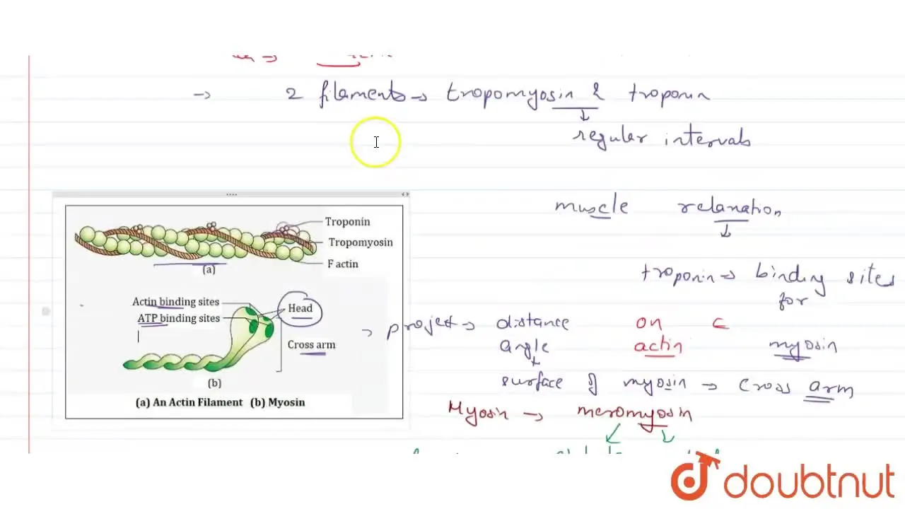
Each actin filaments is having two forms Monomer G-actin and polymeric F-actin.
G-actin is presence of Mg++ forms polymeric filamentous F-actin.
Tropomyosin also run close to the .F. actin throughout its length. br A complex protein troponin is distributed at regular intervals on the tropomyosin.
In a resting state a sub unit of troponin masks the active binding sites of myosin on the actin filaments.
(a) Troponin I TpI(Inhibitor location)
(b) Troponin T TpT(Tropomyosin location)
(c) Troponin C TpC(Ca++ joining location)

Each myosin filaments is formed of polymerised protein. They are present only in A disc
many monomeric proteins called Meromyosins constitute one tick filament.

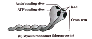
Each meromyosins has two important parts, a globular head with a short arm and a tail, the former being called the heavy meromyosin (HMM) and the latter, the light meromyosin (LMM).
The HMM component, i.e, the head and short arm projects outwards at regular distance and angle from each other from the surface of a polymerised myosin filament and is known as cross arm.
The globular head is an active ATPase enzyme and has binding sites for ATP and active sites for actin.
|
Updated on:21/07/2023
Related Playlists
- BOOK - KUMAR PRAKASHANCHAPTER - LOCOMOTION AND MOVEMENT EXERCISE - SECTION B (Difference /Scientific Reasons)7videos
LOCOMOTION AND MOVEMENT
- BOOK - KUMAR PRAKASHANCHAPTER - LOCOMOTION AND MOVEMENT EXERCISE - SECTION -C (Defination /Explanation -terms /Location -Function /Full Name )17videos
LOCOMOTION AND MOVEMENT
- BOOK - KUMAR PRAKASHANCHAPTER - DIGESTION AND ABSORPTIONEXERCISE - Question from Module (Question Paper)11videos
DIGESTION AND ABSORPTION
- BOOK - KUMAR PRAKASHANCHAPTER - MINERAL NUTRITIONEXERCISE - QUESTIONS FOR MODULE (IMPORTANT MCQ FOR NEET)4videos
MINERAL NUTRITION
Similar Questions
Pectoral girdle and pelvic girdle
07:51View SolutionSkletal muscles
01:36View SolutionWhy are villi present in the intestine and not in the stomach ?
01:22View SolutionWhich enzyme is essential for the formation of nitrogen?
01:56View SolutionStructure Of Contractile Proteins
01:37View SolutionContractile proteins are found in
03:01View Solutionwhich of the following is a contractile protein ?
03:17View SolutionContractile protein is
View SolutionThe contractile protein in the human body is
01:39View SolutionThe major contractile proteins of muscles is/are
36:16View SolutionWrite a note on the structure of contractille protein.
02:00View SolutionContractile protein is -
02:32View Solutionसंकुचनशील प्रोटीन है -
05:17View SolutionStructure Of Contractile Proteins | Mechanism Of Muscle Contration | Part 2
01:51View SolutionExplain the structure of contractile proteins.
00:59View Solution


