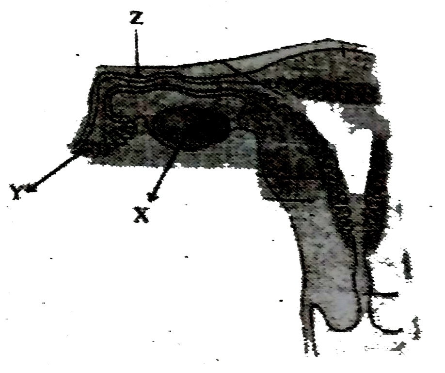
The diagram above shows a part of the human female reproductive system.
(a) Name the gamete cells that would be present in 'X' if taken from a newborn baby.
(b) Name 'Y' and write its function.
(c) Name 'Z' and write the events that take · place here.
(a) X→ Ovary-The gamete cell is Primary Oocyte.
(b) Y→Fimbriae.
Function of Fimbriae→The edge of the in fundibulum possesses finger-like projections called fimbriae, which help in collection of ovum after ovulation.
(c) Z→Ampullary is the mic Function
Ampullary Isthmic Junction → It is the last part of oviduct having a narrow lumen which joins the uterus and mitosis occurs in zygote in the isthmus.
Topper's Solved these Questions
- XII BOARDSBOOK - XII BOARD PREVIOUS YEAR PAPER ENGLISHCHAPTER - XII BOARDSEXERCISE - SET-I Section D
1 Videos
- XII BOARDSBOOK - XII BOARD PREVIOUS YEAR PAPER ENGLISHCHAPTER - XII BOARDSEXERCISE - SET-I Section E
6 Videos
- XII BOARDSBOOK - XII BOARD PREVIOUS YEAR PAPER ENGLISHCHAPTER - XII BOARDSEXERCISE - SET-I Section B
6 Videos
- SAMPLE PAPER 2019BOOK - XII BOARD PREVIOUS YEAR PAPER ENGLISHCHAPTER - SAMPLE PAPER 2019EXERCISE - Section E Part B
3 Videos
Similar Questions
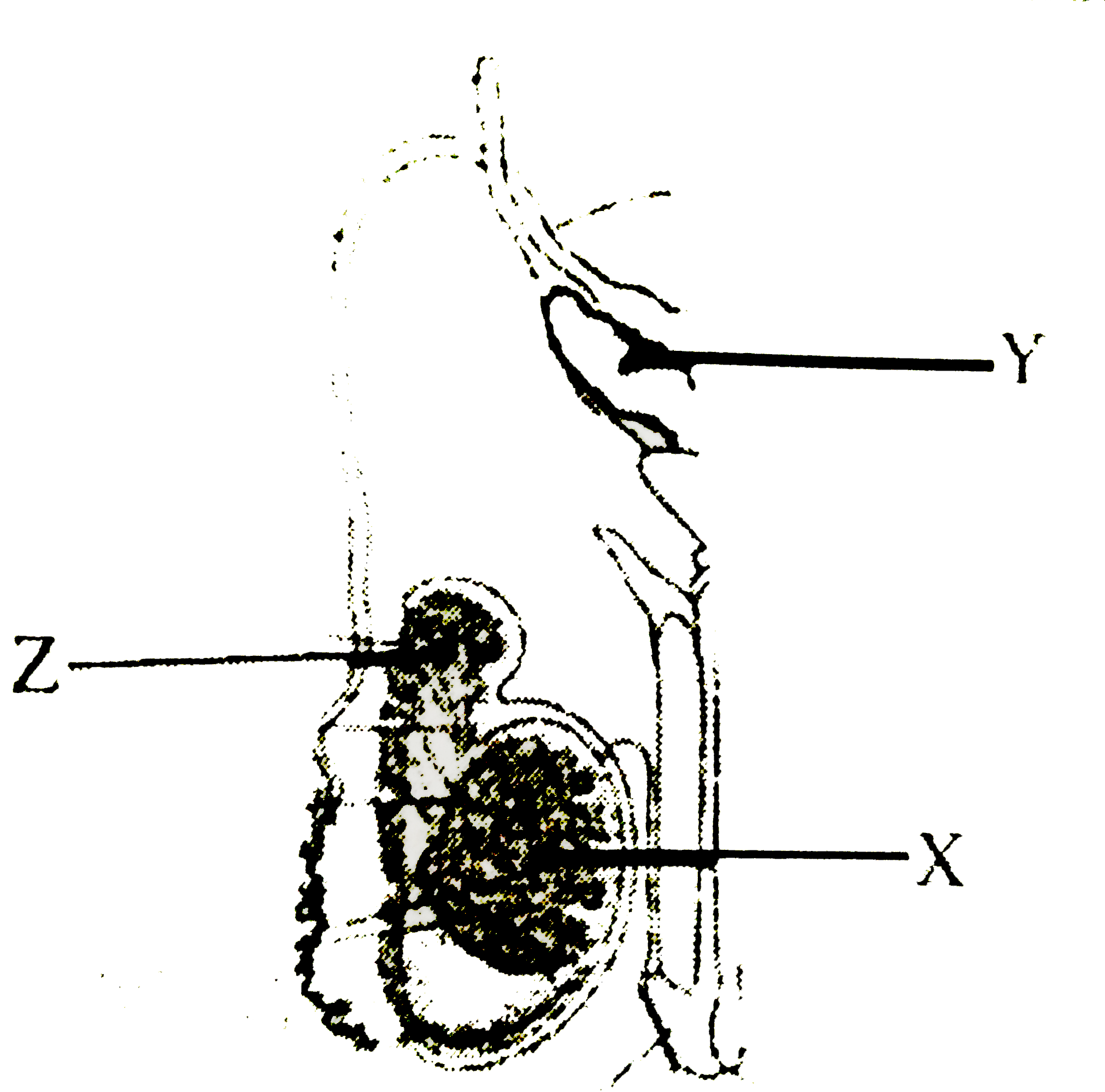
The above diagrams shows human male reproductive system (one side only )
(a) Identify 'X' and write its location in the body .
(b) Name the accessory gland 'Y' and its reaction.
(c) Name and state the function of 'Z'.
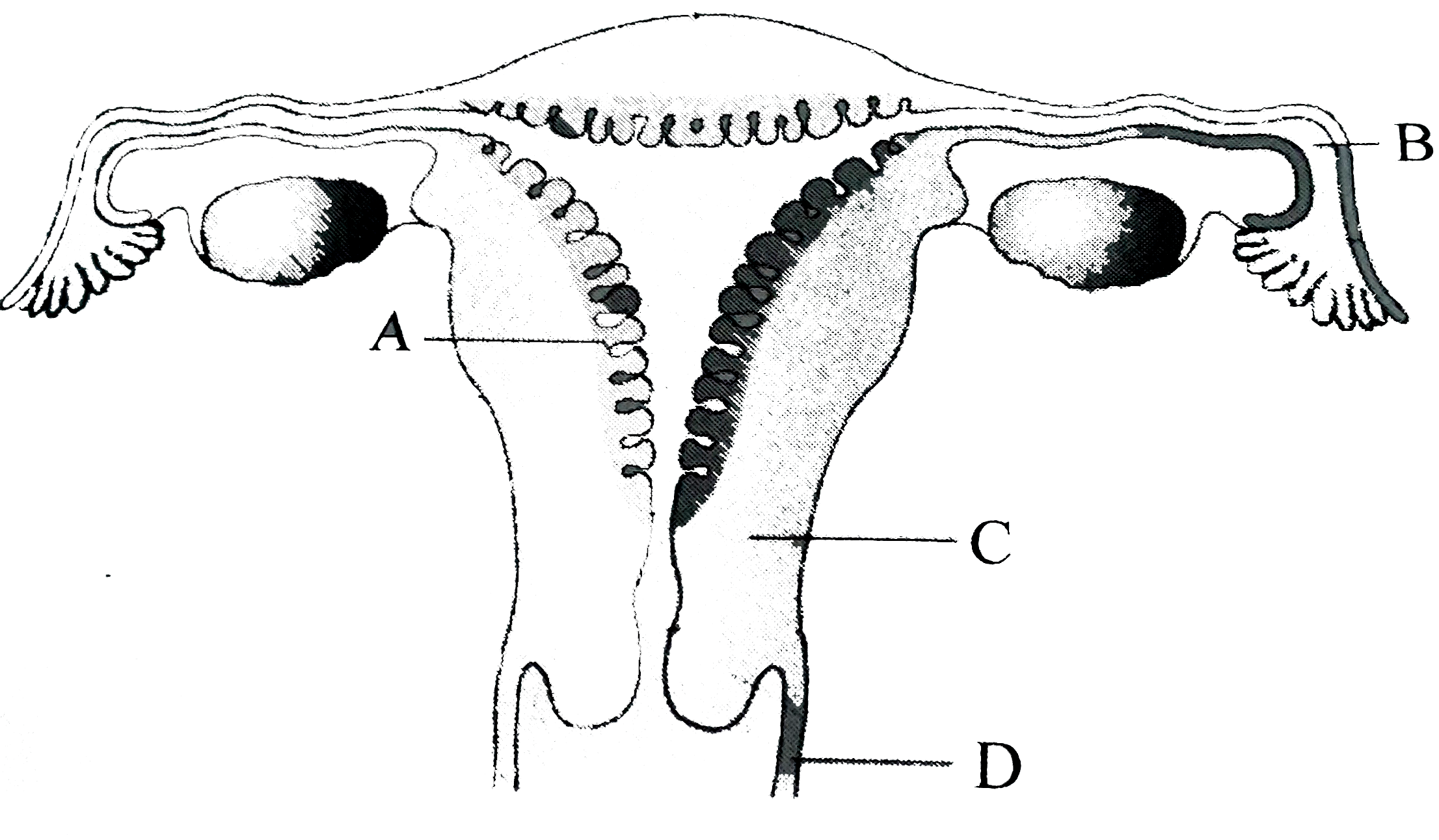
View SolutionThe given diagram shows a human female 's reproductive system. Select the part (labelled as A, B, C & D) where semen is released into the female by the penis during copulation
View Solution
Knowledge Check
The given diagram shows a duct system of liver, gall bladder and pancrease. Write the names of ducts from A to D.
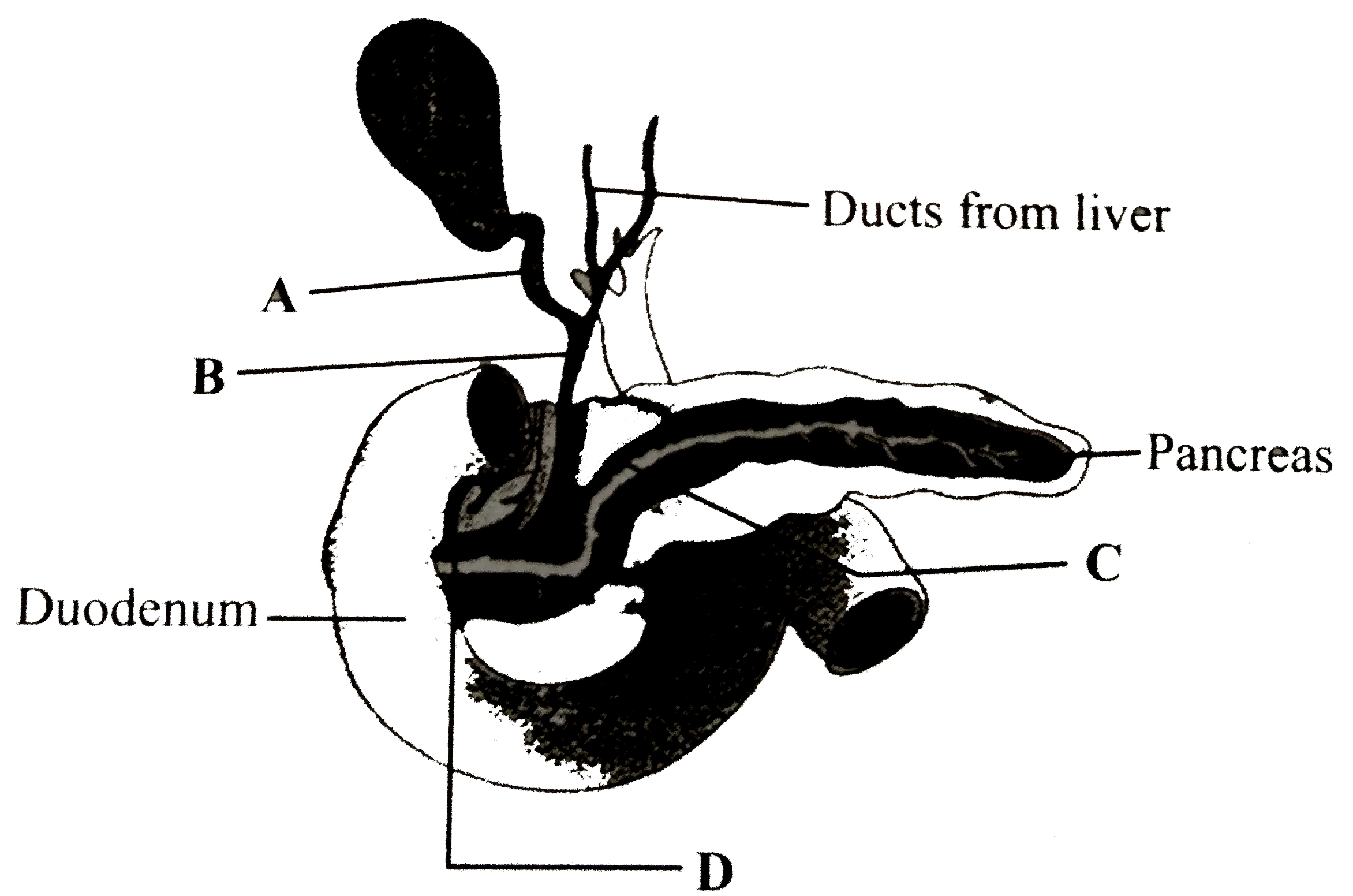 AA-Cystic duct,B-Common bile duct, C-Pancreatic duct, D-Hepatopancreatic ductBA-Common bile duct, B-Cystic duct, C-Pancreatic duct,D-Hepatopancreatic ductCA-Cystic duct, B-Bile duct, C-Hepatopancreatic duct, D-Pancreatic ductDA-Cystic duct, B-Panecreatic duct, C-Common bile duct, D-Hepatopancreatic duct
Given here is a section of the female reproductive system in humans.
a. Name the parts labelled 1 to 4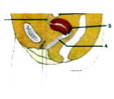
b. Name the part where fertilisation occurs in the female reproductive system.
View SolutionName the respective part of human female reproductive system:
(i) that produces eggs.
(ii) where fusion of egg and sperm take place, and
(iii) where zygote implanted.
(b) Describe in brief what happen to the zygote after it gets implanted.
View Solution(a) Write the function of following parts in human female reproductive system :
(i) Ovary , (ii) Oviduct , (iii) Uterus
(b) Describe in brief the structure and function of placenta .
View SolutionThe diagram given below represents two reproductive cells A and B. Study the same and then answer the questions that follow:
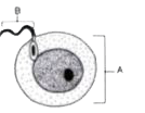
(i) Identify the reproductive cells A and B.
(ii) Name the specific part of the reproductive system where the above cells are produced
(iii) Where in the female reproductive system do these cells unite ?
(iv) Name the main hormones secreted by the (1) ovary (2) testes.
(v) Name an accessory gland found in the male reproductive system and state the function of its secretion.
View SolutionStudy the diagram given alongside showing a plant cell kept in a strong sugar solution.
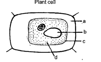
Name the parts a, b, c, and d.
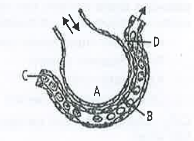
View SolutionThe figure given below shows a small part of human lung where exchange of gases takes place in which one of the options given below the one part A,B,C or D is correctly identified along with its function
View SolutionGiven below is the outline of the male reproductive system :
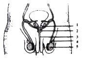
(i) Name the parts labelled 1 to 5.
(ii) State the functions of the parts labelled 1 and 4.
(iii) Name the cells of part 5 that produce testosterone.
(iv) Why is the structure 5 present outside the body in the scrotal sacs ?
(v) What is semen ?
(vi) Give the exact location of prostate gland.
View Solution
