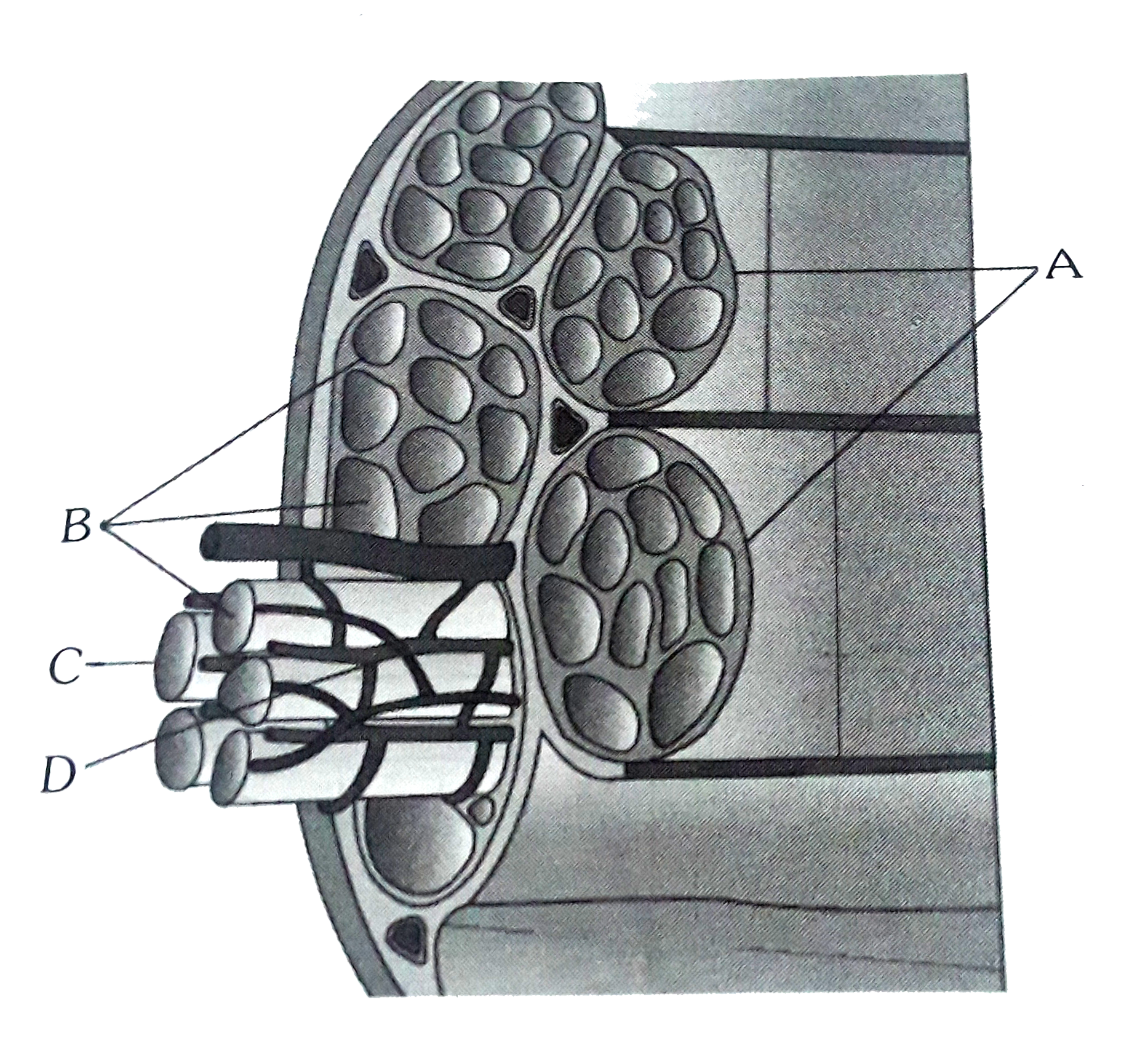
Label the following parts in the diagram of eye :
(a) Aqueous chamber
(b) Cornea
(c) Lens
(d) Retina
(e) Vitreous chamber
(f) Blind spot
To label the parts of the eye in the diagram, follow these steps:
1. Identify the Aqueous Chamber:
- Look for the front part of the eye, which is filled with a clear fluid. This area is known as the aqueous chamber. It is located between the cornea and the lens.
- Label: Aqueous Chamber
2. Locate the Cornea:
- The cornea is the transparent front part of the eye that covers the iris, pupil, and anterior chamber. It is the first structure that light passes through and is responsible for refracting light.
- Label: Cornea
3. Find the Lens:
- The lens is situated behind the iris and the pupil. It is a transparent structure that focuses light onto the retina. It can change shape to help focus on objects at various distances.
- Label: Lens
4. Identify the Retina:
- The retina is located at the back of the eye. It is a thin layer of tissue that contains photoreceptor cells (rods and cones) and is responsible for converting light into neural signals.
- Label: Retina
5. Locate the Vitreous Chamber:
- The vitreous chamber is the large space behind the lens and in front of the retina. It is filled with a gel-like substance called vitreous humor that helps maintain the shape of the eye.
- Label: Vitreous Chamber
6. Identify the Blind Spot:
- The blind spot is located where the optic nerve exits the eye. This area does not contain photoreceptor cells, which is why it cannot detect light, making it a "blind" spot.
- Label: Blind Spot
- Look for the front part of the eye, which is filled with a clear fluid. This area is known as the aqueous chamber. It is located between the cornea and the lens.
- Label: Aqueous Chamber
- The cornea is the transparent front part of the eye that covers the iris, pupil, and anterior chamber. It is the first structure that light passes through and is responsible for refracting light.
- Label: Cornea
- The lens is situated behind the iris and the pupil. It is a transparent structure that focuses light onto the retina. It can change shape to help focus on objects at various distances.
- Label: Lens
- The retina is located at the back of the eye. It is a thin layer of tissue that contains photoreceptor cells (rods and cones) and is responsible for converting light into neural signals.
- Label: Retina
- The vitreous chamber is the large space behind the lens and in front of the retina. It is filled with a gel-like substance called vitreous humor that helps maintain the shape of the eye.
- Label: Vitreous Chamber
- The blind spot is located where the optic nerve exits the eye. This area does not contain photoreceptor cells, which is why it cannot detect light, making it a "blind" spot.
- Label: Blind Spot
Topper's Solved these Questions
- NEURAL CONTROL AND COORDINATIONBOOK - MODERN PUBLICATIONCHAPTER - NEURAL CONTROL AND COORDINATIONEXERCISE - NCERT Exemplar Problems (D. LONG ANSWER TYPE QUESTIONS)
3 Videos
- NEURAL CONTROL AND COORDINATIONBOOK - MODERN PUBLICATIONCHAPTER - NEURAL CONTROL AND COORDINATIONEXERCISE - Higher Order Thinking Skills & Brain Twisting (Very Short Answer Questions)
13 Videos
- NEURAL CONTROL AND COORDINATIONBOOK - MODERN PUBLICATIONCHAPTER - NEURAL CONTROL AND COORDINATIONEXERCISE - NCERT Exemplar Problems (B. VERY SHORT ANSWER TYPE QUESTIONS)
12 Videos
- MORPHOLOGY OF FLOWERING PLANTS BOOK - MODERN PUBLICATIONCHAPTER - MORPHOLOGY OF FLOWERING PLANTS EXERCISE - CASE BASED SHORT ANSWER TYPE QUESTIONS
2 Videos
- PHOTOSYNTHESIS IN HIGHER PLANTSBOOK - MODERN PUBLICATIONCHAPTER - PHOTOSYNTHESIS IN HIGHER PLANTSEXERCISE - CHAPTER PRACTICE TEST
9 Videos
Similar Questions
Knowledge Check
See the following diagram and identify A,B,C,D,E,F and G
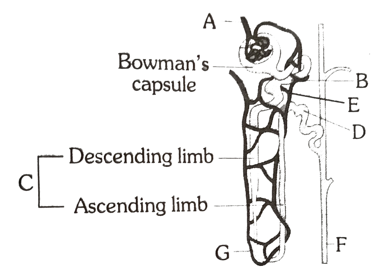 AA-Afferent arteriole, B-Henle' s loop, C-Collecting duct, D-PCT, E-DCT, F-Pertibular capillaries, G- vasa rectaBA-afferent arteriole, B-Peritubular capillaries, C-Helne's loop, D-DCT, E-PCT, F-Collecting duct, G-vasa rectaCA-Afferent arteriole, B- PCT, C-Henle's loop, D-DCT, E-Peritublar capillaries, F-Collecting duct, G-vasa rectaDA-Afferent arteriole, B-Proximal convoluted tubule, C-Henle's loop, D-Distal convoluted tubule, E-Peritbular capillaries, F-Collecting duct, G-vasa recta
The following is the diagram of T.S of anther. Identify the parts labelled A, B and C
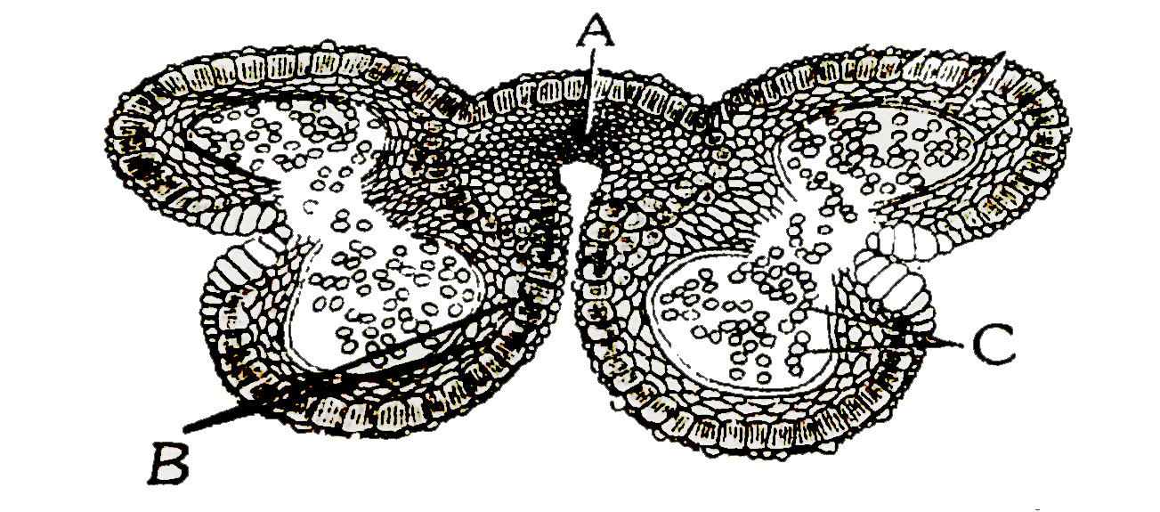 AA-Connective tissue, B-Endothecium, C- Pollen grainBA- Endothecium, B- Connective tissue, C- Pollen grainCA-Pollen grain, B- Connective tissue, C- EndotheciumDA- Endothecium, B- Pollen grain, C- Connective tissue
The following is the diagram of T.S. of anther. Identify the parts labeled A, B and C.
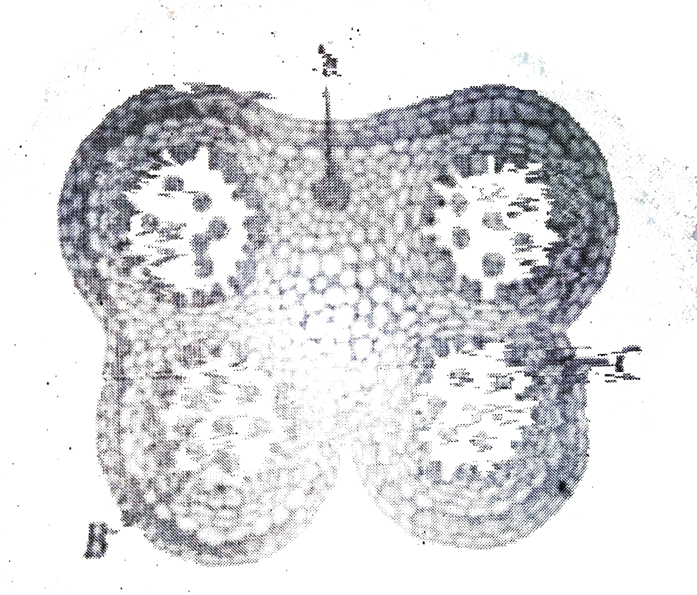 AA-Connective tissue, B-Pollen grains, C-EndotheciumBA-Endothecium, B-Connective tissue, C-Pollen grainsCA-Pollen grains, B-Connective tissue, C-EndotheciumDA-Endothecium, B-Pollen grains, C-Conncetive tissue
Draw labelled diagrams of the following :
(a) Neuron
(b) Brain
(c) Eye
(d) Ear
View SolutionExplain the function of the following parts fo human eye and brain.
(a) Caronea (b) Iris ( c) Pupil (d) Lens ( e) Reatina and optic nerves (f) Brain
View SolutionIn the following diagram of section of a human heart, label a, b, c, d, e and f.
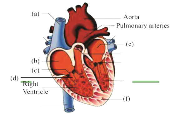
View SolutionDraw the following diagrams and label their parts:
(a) Stamen
(b) Pistil
View SolutionLabel the parts of the following diagram of the human eye and state the functions of those parts.
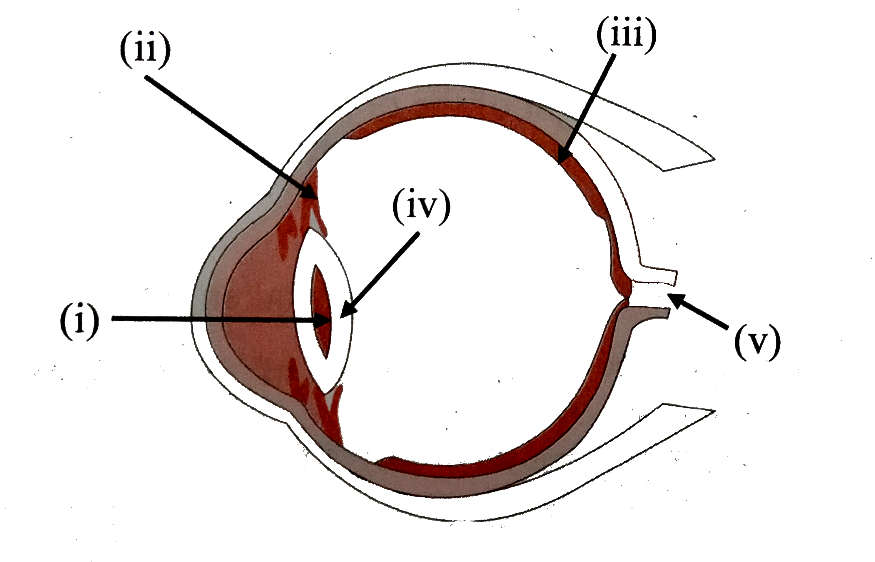
View Solution


