Which of the following figure shows the mandibles of cockroach ?
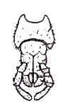
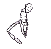
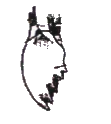
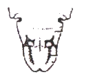
The correct Answer is:C
To determine which figure shows the mandibles of a cockroach, follow these steps:
1. Understand the Anatomy of Cockroach Mouth Parts:
- Cockroaches have a complex mouth structure that includes several parts: labrum (upper lip), mandibles, maxillae, labium (lower lip), and hypopharynx (tongue).
2. Identify the Function of Mandibles:
- The mandibles are primarily responsible for biting, chewing, and grasping food. They are strong and pointed at the ends, which aids in these functions.
3. Examine the Options Provided:
- Look at each figure provided in the options. Identify which figure corresponds to the mandibles based on their shape and function.
4. Analyze Each Option:
- Option 1: This figure represents the labium (lower lip), which is incorrect.
- Option 2: This option does not match the mandibles either.
- Option 3: This figure shows the mandibles, characterized by their pointed ends, which is correct.
- Option 4: This figure represents the hypopharynx (tongue), which is also incorrect.
5. Select the Correct Answer:
- Based on the analysis, the correct answer is Option 3, as it accurately depicts the mandibles of a cockroach.
Final Answer:
The figure that shows the mandibles of a cockroach is Option 3.
---
- Cockroaches have a complex mouth structure that includes several parts: labrum (upper lip), mandibles, maxillae, labium (lower lip), and hypopharynx (tongue).
2. Identify the Function of Mandibles:
- The mandibles are primarily responsible for biting, chewing, and grasping food. They are strong and pointed at the ends, which aids in these functions.
- Look at each figure provided in the options. Identify which figure corresponds to the mandibles based on their shape and function.
- Option 1: This figure represents the labium (lower lip), which is incorrect.
- Option 2: This option does not match the mandibles either.
- Option 3: This figure shows the mandibles, characterized by their pointed ends, which is correct.
- Option 4: This figure represents the hypopharynx (tongue), which is also incorrect.
- Based on the analysis, the correct answer is Option 3, as it accurately depicts the mandibles of a cockroach.
The figure that shows the mandibles of a cockroach is Option 3.
Similar Questions
Knowledge Check
- Question 1 - Select One
Mandibles of cockroach are :
AShort with grinding teethBLong and pointedCshort without teethDlong and coiled - Question 2 - Select One
Mandibles of cockroach are :
ALong and pointedBShort without teethCPerforated, syringe likeDWith grinding and incising regions - Question 3 - Select One
Which of the following figures shows tubectomy ?
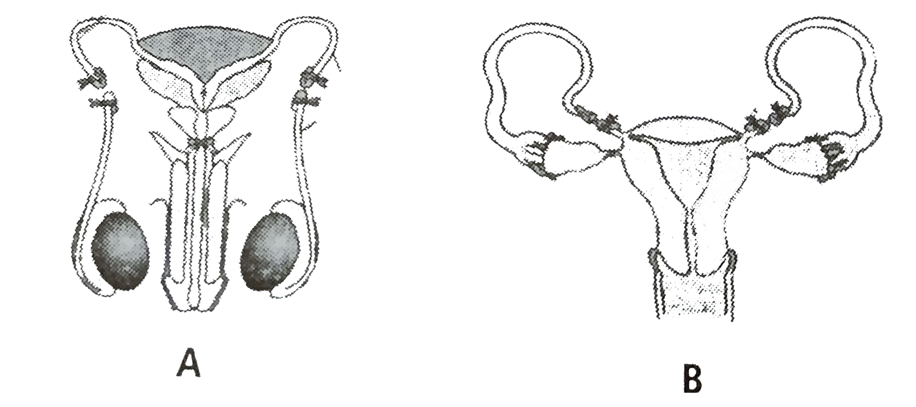 AA onlyBB onlyCEither A or BDNone of these
The following figures show
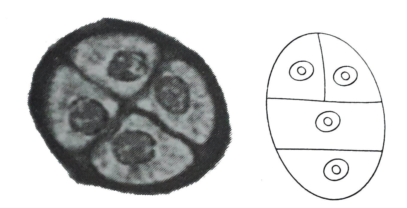 View Solution
The following figure shows the process of
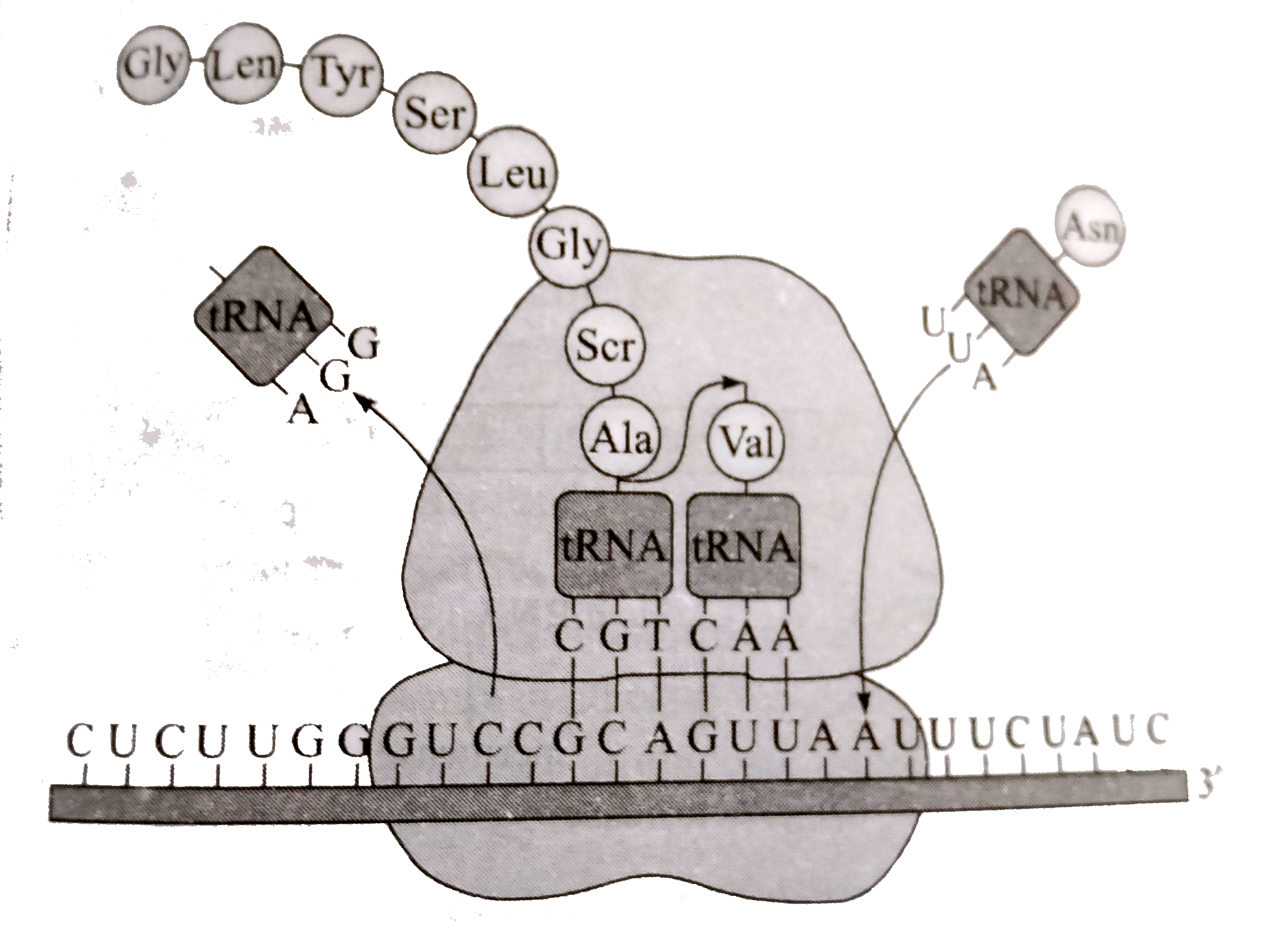 View Solution
The following figure shows the phenomenon of
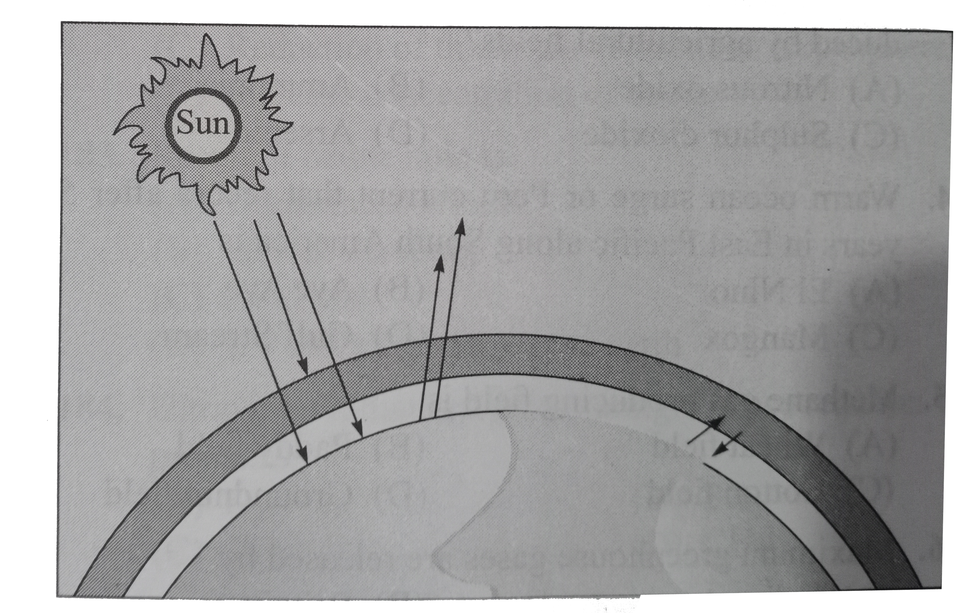 View Solution
The following figure shows the external features of cockroach with few structure labelled as A,B, C, D & E.
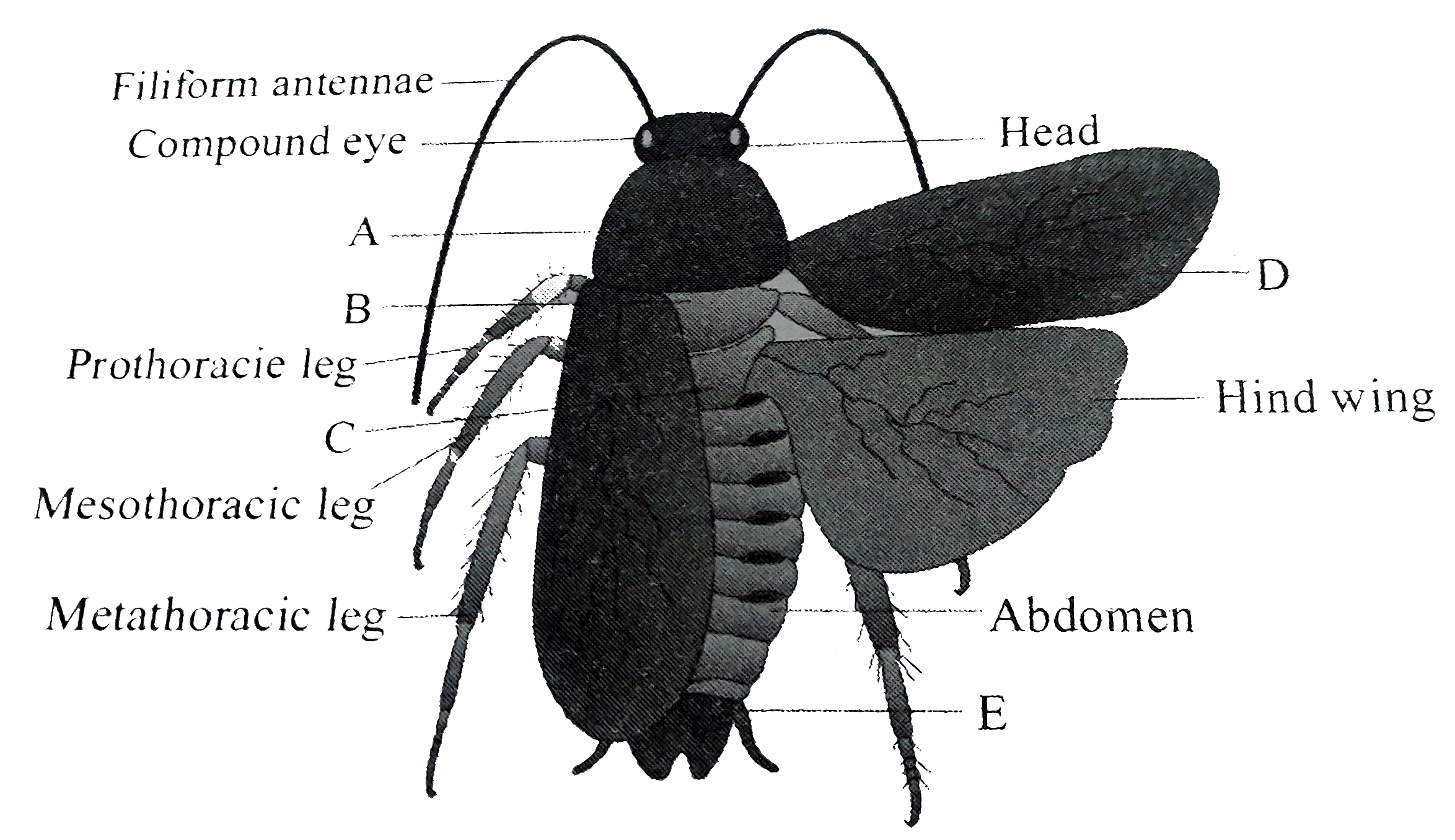
Identify A to EView SolutionWhich of the following figures show(s) the bonding in diborane ?
View Solution

